What HEPES reveals about cultivating with light
When the chemical used to control an experiment becomes part of the experiment.
A cyanobacterial growth recipe looks modular. BG-11 supplies nutrients, HEPES holds the pH steady, light drives photosynthesis, the cyanobacterium grows. Each ingredient has one job.
The buffer in many cyanobacteria and algae recipes. It is added to keep the pH from drifting as the cells feed and change the water around them.
The organism never encounters the word buffer. It encounters a molecule in a chemical environment, and when the lights come on that environment changes.
Illuminated medium containing HEPES generates hydrogen peroxide. That was reported in 1985, in a paper asking why cells kept dying in light-exposed medium (Zigler et al., 1985). How much depends on the buffer’s amine. Tertiary amines produce the most and primary amines almost none, which is why Tris sits on the no-buffer baseline and HEPES sits far above it (Liu et al., 2023).
Whether that matters is a biological question, not a chemical one.
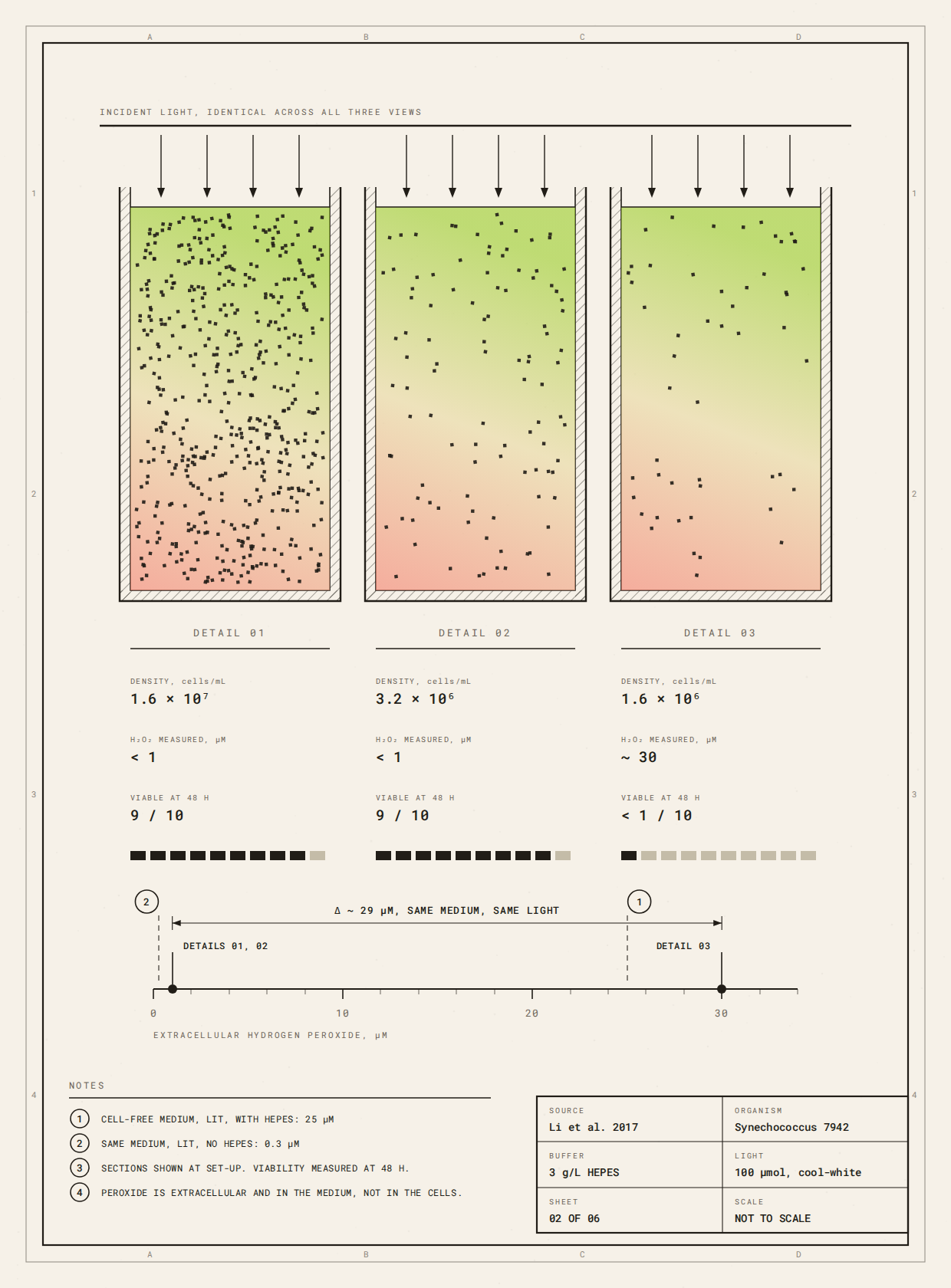
Fig. 1. Cell number decides whether the buffer matters. One medium, one light level, three starting densities. Lit with no cells in it, this medium reaches about 25 µM hydrogen peroxide. The two dense cultures hold it under 1 µM and nine in ten cells are alive at 48 hours. The dilute culture sits near 30 µM and fewer than one in ten survive. Sections drawn at set-up. Redrawn from Li et al. (2017).
Prochlorococcus is the clearest case. It lacks catalase, and neighbouring heterotrophs keep it alive by pulling peroxide out of the shared water (Morris et al., 2011). In illuminated seawater buffered with HEPES, axenic cultures died. They lived if a helper bacterium was present, or if the buffer was swapped. Cutting the HEPES until it made 75% less peroxide did not save them.
Losing your own defences only counts as a deficiency if you are alone.
Cell density settles the same question in the lab. Li and colleagues found dense cyanobacterial cultures untroubled by the peroxide in their medium while dilute ones died within two days (Li et al., 2017). Their fix was another organism. In their hands, and in Morris’s, a living partner outperformed purified catalase.
Sometimes the way to hold a system steady is to put something else living into it, rather than take a variable out.
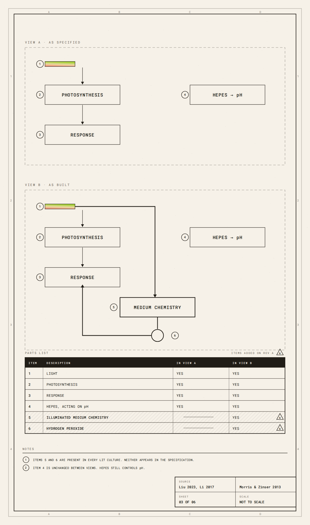
Fig. 2. The system as specified, and the system as built. Both views run the same light through the same culture, and in both the buffer holds pH exactly as intended. The built version carries two parts the specification never lists: the medium’s own photochemistry, and the peroxide it makes, which feeds back into the thing being measured. Nothing here is broken. The parts list is incomplete.
This is where it reaches design. Designers change cell loading, thickness, porosity, geometry and illumination. Those read as fabrication variables. For a living system they are environmental ones. Printed microalgal hydrogels are described as habitats that shape light and mass transport (Dawiec-Liśniewska et al., 2026). Alginate scaffolds swap ions with the medium and loosen, which changes gas exchange and then carbon fixation (Levä et al., 2023). Photosynthetic beads lose pigment where light cannot reach the cells (Armaly et al., 2026).
Change the geometry and you have changed what the buffer does.
Which is why “HEPES-buffered” does not specify an experiment. Published methods give concentration but not spectrum, or density but not whether peroxide was measured (Tóth et al., 2022; Wang et al., 2026; Morris & Zinser, 2013). Nothing in them is false. The record is not sufficient to tell whether this chemistry was running.
Biodesign teaching is already close to the right posture. Living Pigments asks students to treat organisms as collaborators and to respond to biological variability rather than suppress it (Nerlich et al., 2026). The HEPES case adds one step. Ask how the environment changes the organism, then ask how it changes the things you introduced to control the organism.
The first check is cheap. Put your medium under your own light, with no cells in it, and measure peroxide over your own timescale. One assay, no biology, and it tells you whether there is anything worth chasing.
Short version of Reading 05. The HEPES definition is lifted verbatim from the Biodesign Academy glossary. Full Reading and Members’ Teaching Notes in the Library.
Reading 05, paper by paper
This is the short version. The full Reading works through the evidence paper by paper, with page-level sourcing, six plates, and the cases where the effect does not appear.
Members also get the Teaching Notes: four questions for auditing any control in a protocol, a printable audit card, candidate second pathways in fabricated living systems, and the reagent record a machine would need before it could reason about a protocol.
Armaly, P., Shachor, G., Berger, Y., Iliassafov, L., Rosenblau, K., Kashi, Y., & Barath, S. (2026). Biodesign approaches for photosynthetic microbial applications in architecture. Biotechnology Design. doi: 10.1017/S2977905726100961.
Dawiec-Liśniewska, A., et al. (2026). Biodesign of microalgae-laden engineered living materials via 3D bioprinting: a roadmap. Biotechnology Design. doi: 10.1017/S2977905726100869.
Levä, T., et al. (2023). Mapping nanocellulose- and alginate-based photosynthetic cell factory scaffolds: interlinking porosity, wet strength, and gas exchange. Biomacromolecules, 24, 3484-3497. doi: 10.1021/acs.biomac.3c00261.
Li, T., et al. (2017). Mimicking lichens: incorporation of yeast strains together with sucrose-secreting cyanobacteria improves survival, growth, ROS removal, and lipid production in a stable mutualistic co-culture production platform. Biotechnology for Biofuels, 10, 55. doi: 10.1186/s13068-017-0736-x.
Liu, P., et al. (2023). Zwitterionic betaines over HEPES as the new generation biocompatible pH buffers for cell culture. Bioactive Materials, 24, 376-386. doi: 10.1016/j.bioactmat.2022.12.028.
Morris, J. J., Johnson, Z. I., Szul, M. J., Keller, M., & Zinser, E. R. (2011). Dependence of the cyanobacterium Prochlorococcus on hydrogen peroxide scavenging microbes for growth at the ocean’s surface. PLoS ONE, 6(2), e16805. doi: 10.1371/journal.pone.0016805.
Morris, J. J., & Zinser, E. R. (2013). Continuous hydrogen peroxide production by organic buffers in phytoplankton culture media. Journal of Phycology, 49(6), 1223-1228. doi: 10.1111/jpy.12123.
Nerlich, P., Archer, L., & Morgado Diniz, N. (2026). Teaching biodesign through algal experimentation: methods for living-system prototyping. Biotechnology Design, 4, e32, 1-15. doi: 10.1017/S2977905726100328.
Tóth, G. S., et al. (2022). Photosynthetically produced sucrose by immobilized Synechocystis sp. PCC 6803 drives biotransformation in E. coli. Biotechnology for Biofuels and Bioproducts, 15, 146. doi: 10.1186/s13068-022-02248-1.
Wang, M., et al. (2026). Co-culture of mammalian cells and photosynthetic microorganisms for oxygen supply in engineered tissues. Cell Proliferation, advance online publication, e70224. doi: 10.1111/cpr.70224.
Zigler, J. S., Jr., et al. (1985). Analysis of the cytotoxic effects of light-exposed HEPES-containing culture medium. In Vitro Cellular & Developmental Biology, 21, 282-287. doi: 10.1007/BF02620943.
From the Molecule Up, by Biodesign Academy. Read one living material with care, as a relation you are joining, then meet the next on its own terms.
