What HEPES reveals about cultivating with light
When the chemical used to control an experiment becomes part of the experiment.
A cyanobacterial growth recipe looks reassuringly modular. BG-11 supplies nutrients, HEPES keeps the pH steady, light drives photosynthesis, the cyanobacterium grows.
Describing protocols this way is useful. Without functional labels even a simple recipe is hard to read.
But the organism doesn’t encounter the word buffer. It encounters a molecule in a chemical environment, and when the lights come on that environment can change.
HEPES opens onto a broader problem: control interference. Something introduced to hold one part of an experiment steady can also change another part the organism cares about.
The control still works. It just doesn’t stay outside the experiment.
HEPES is doing its job
HEPES is one of the Good’s buffers. Cells shift the pH around them, and a buffer reduces that drift. So HEPES → pH control is a fair description.
The description is fine. The trouble is what disappears once the label is assigned.
Researchers have known this for decades. In 1985 Zigler and colleagues left HEPES-containing culture medium under visible light and found it had turned cytotoxic. HEPES and riboflavin were enough on their own to produce the effect, and hydrogen peroxide accounted for most, though not all, of the inhibition (Zigler et al., 1985).
Recent work explains how. Riboflavin acts as a photosensitiser, producing short-lived reactive oxygen species under visible light. The tertiary amine chemistry of HEPES then converts much of that transient chemistry into hydrogen peroxide, which lasts.
The same study tested 7 further Good’s buffers. Tertiary amines produced 91 to 157 µM peroxide. Secondary amines produced around 60. Tris, a primary amine, produced none that could be detected (Liu et al., 2023).
The amine is doing the work, and which kind decides how much.
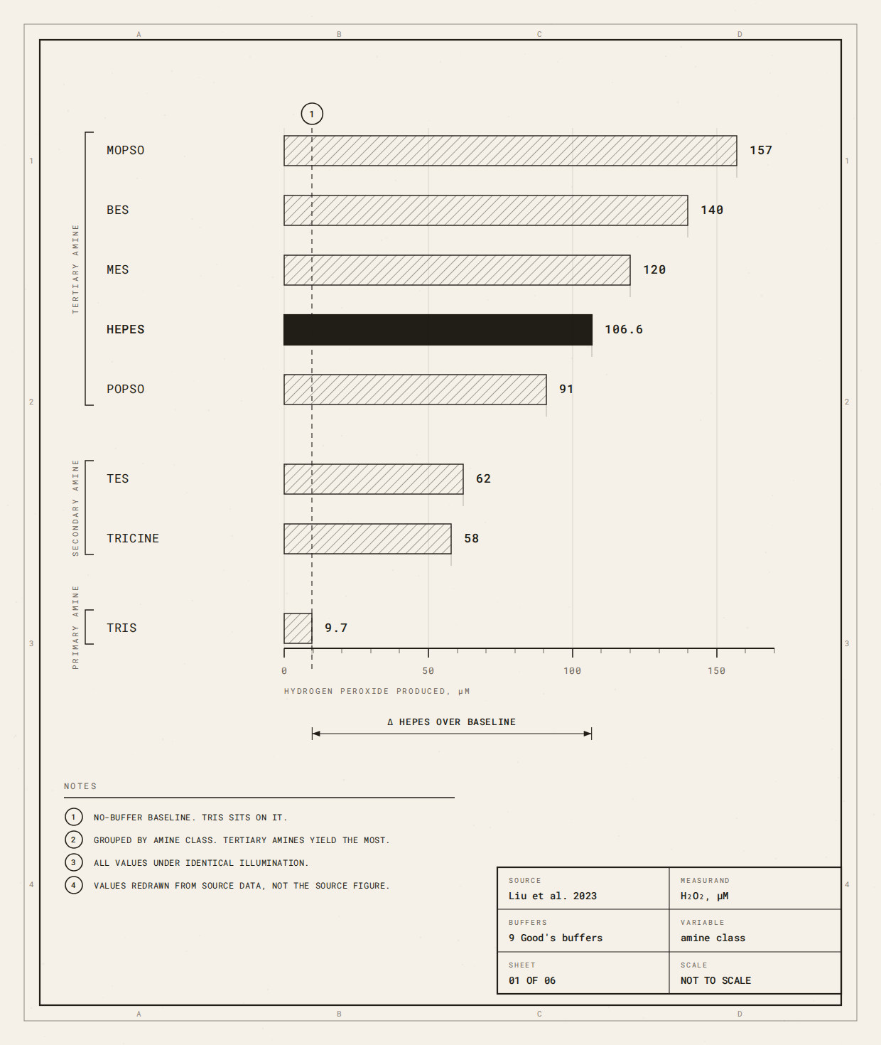
Fig. 1: Which amine, and how much. Eight Good’s buffers held under the same light, in the same medium, for the same time. The 4 tertiary amines produced 91 to 157 µM hydrogen peroxide. The 2 secondary amines produced around 60. Tris, a primary amine, produced none that could be detected. Values from Liu et al. (2023). One medium, one light source, one exposure. Buffering range differs across these molecules and has to be checked separately.
This isn’t HEPES + light = peroxide under every condition. Medium composition matters (Morris & Zinser, 2013). Photosensitisers matter (Liu et al., 2023). Oxygen has to be present at all (Zigler et al., 1985; Morris & Zinser, 2013). So do concentration, light intensity and duration (Morris & Zinser, 2013; Liu et al., 2023).
HEPES stabilises pH. Under some illuminated conditions it also alters the reactive-oxygen environment.
The same substance occupies both pathways, and that’s where control interference begins.
When the background becomes biological
Chemistry becomes biologically important when its products build up faster than the system can clear them.
Prochlorococcus is the clear case. It’s unusually vulnerable to extracellular peroxide, and its genomes lack catalase and other protections common in aerobes. Morris and colleagues showed that neighbouring heterotrophs protect it by pulling peroxide out of the shared water (Morris et al., 2011).
Oxidative tolerance can be a property of the community rather than the cell.
Morris and colleagues push it further. The lineages that moved into the bright surface layer are the ones most sensitive to peroxide, and they shed protective machinery on the way in. Their reading is that the streamlining and the dependence arrived together, as an acquired reliance on the community that clears peroxide for them (Morris et al., 2011).
Losing your own defences only counts as a deficiency if you’re alone.
The same study ran it in reverse. In illuminated seawater with 3.75 mM HEPES, peroxide built up at roughly 0.4 µM per day and axenic Prochlorococcus died. Cultures lived if the helper was present, or if the HEPES was swapped for TAPS, which generates far less peroxide.
Cutting the HEPES until it made 75% less peroxide didn’t save them.
The medium had been built that way on purpose. HEPES was chosen because it produces peroxide steadily under light (Morris et al., 2011).
Morris and Zinser then asked what the medium itself contributed. HEPES produced more peroxide than several other buffers they tested, rising with its concentration, the proportion of natural seawater and the light level. At concentrations commonly used in cultivation it produced enough to stop axenic growth entirely (Morris & Zinser, 2013).
The result is narrower than a verdict on HEPES. A chemical introduced to control pH had become coupled to illumination, and its consequence depended on the organism and the ecology around it.
The buffer had become part of the environment it was meant to stabilise.
Density changes what the buffer means
The clearest example comes from Synechococcus elongatus PCC 7942.
Li and colleagues were building a co-culture of sucrose-producing cyanobacteria and yeast in a medium containing 3 g/L HEPES. It looked unremarkable. Against HEPPSO and TAPS, all 3 buffers supported similar growth and sucrose production while holding pH (Li et al., 2017).
Then they changed the starting density.
Cultures beginning at about 1.6 × 10⁷ or 3.2 × 10⁶ cells per millilitre grew. Those at 1.6 × 10⁶ didn’t, with the living fraction falling below 10% within 2 days before the culture died.
Peroxide tracked the difference. Dense cultures held it below roughly 1 µM, a figure the paper reports without showing. In the dilute culture it climbed towards 30 µM, close to illuminated cell-free medium.
The same medium under the same light, without HEPES in it, stayed flat at around 0.3 µM (Li et al., 2017).
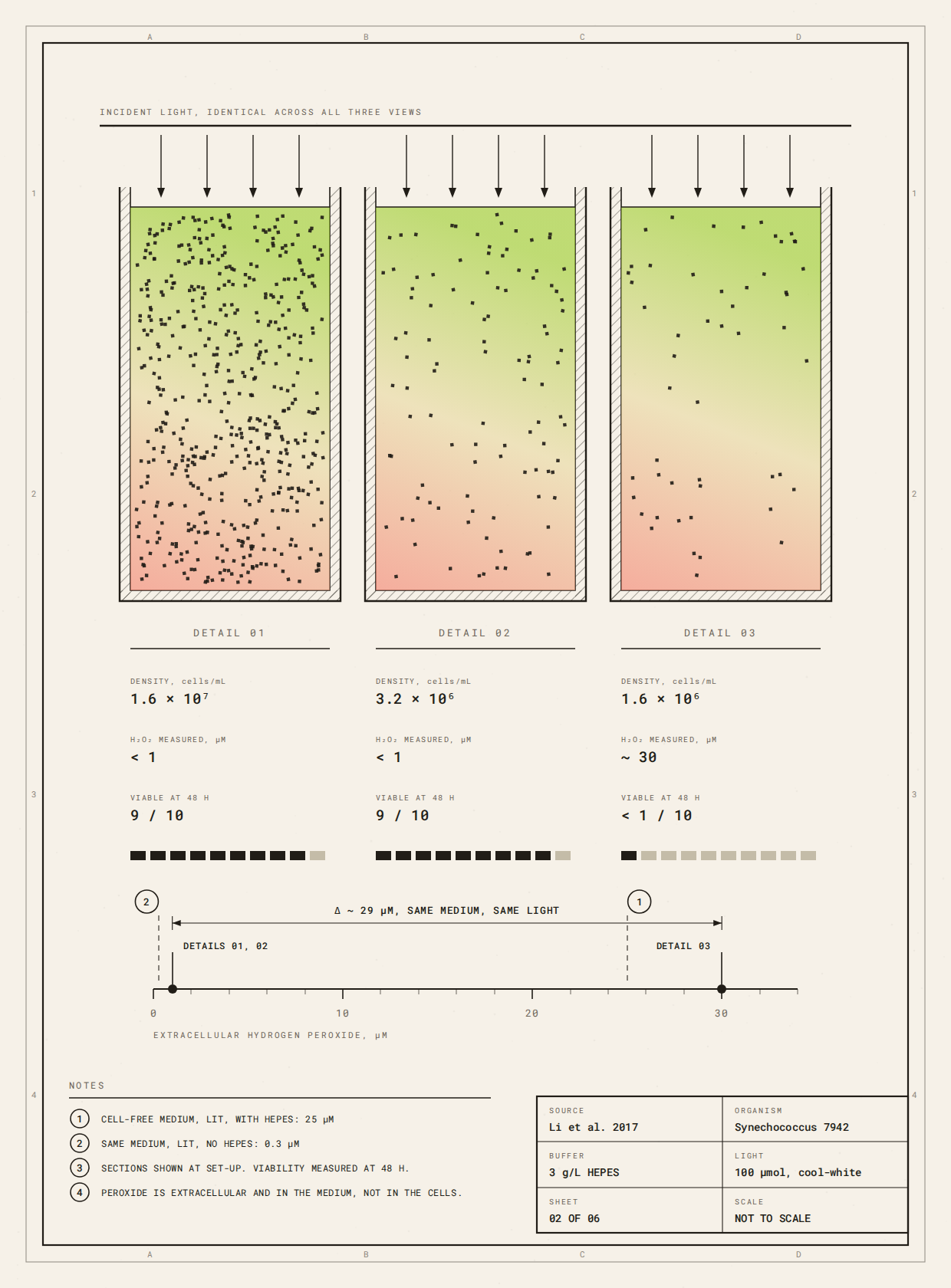
Fig. 2: Same medium, same light, 2 outcomes. Synechococcus elongatus PCC 7942 at 3 starting densities, with reference lines for the medium alone: lit with HEPES in it, and lit without. The 2 denser cultures held extracellular peroxide below roughly 1 µM and grew. The culture started at 1.6 × 10⁶ cells per millilitre climbed towards 30 µM, close to the cell-free line, and fewer than 1 cell in 10 was alive after 2 days. Three measured conditions in one study. No threshold is implied for any other system.
Catalase let the dilute cultures survive. So did the yeast Rhodotorula glutinis, which pulled peroxide out of the water. In dense cultures, where peroxide was never limiting, the yeast beat catalase, so it’s contributing something beyond peroxide removal (Li et al., 2017).
Li and colleagues called the paper Mimicking lichens, and the lichen is the right figure for what they found. Their answer to a chemical problem was another organism, and the organism did more for the cyanobacteria than the enzyme did.
The pattern wasn’t new. Morris and colleagues had shown that Prochlorococcus scavenges external peroxide poorly but can drag it down by mass action once the cells are dense enough, so a culture too dilute to protect itself dies where a thicker one lives (Morris et al., 2011).
HEPES didn’t kill the cyanobacteria. The same environment produced different outcomes because the system’s capacity to handle it changed.
Its apparent neutrality was conditional.
Control interference
Control interference happens when something introduced to hold one part of an experiment steady also changes another part that matters biologically.
HEPES reduces variation in pH. Under some illuminated conditions the medium containing it also affects the oxidative environment (Zigler et al., 1985; Morris & Zinser, 2013; Liu et al., 2023).
The control is still controlling pH, which is what makes it easy to miss.
Testing light intensity, the intended model is light → photosynthesis → response, with HEPES → stable pH sitting outside the causal chain. If illumination also changes the medium’s chemistry, a second pathway appears: light → medium photochemistry → peroxide → response.
Holding the concentration of HEPES constant isn’t the same as holding constant everything HEPES contributes to.
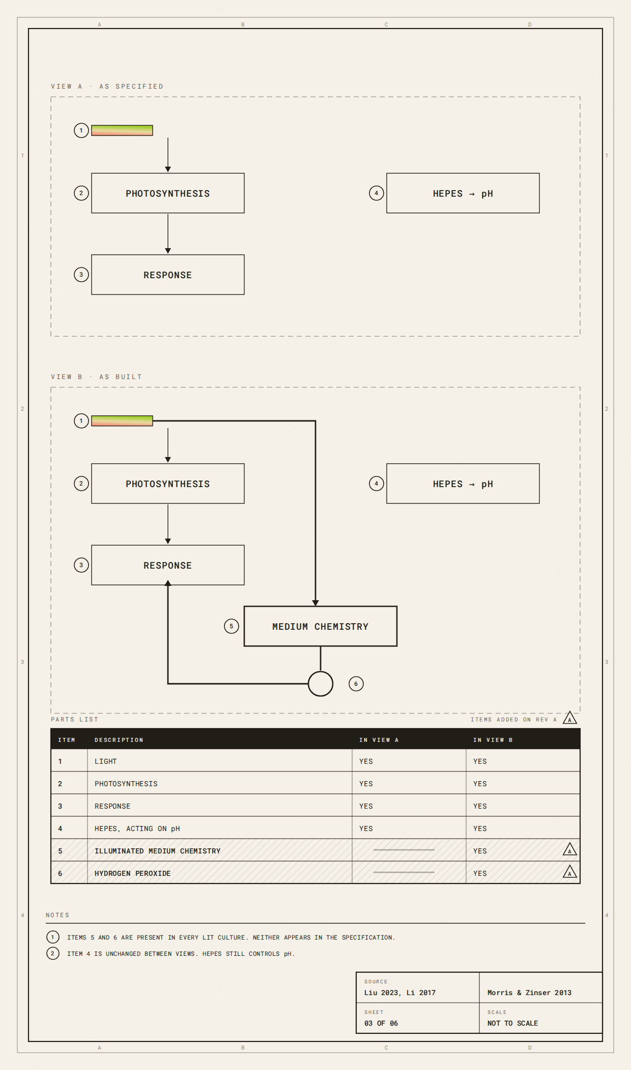
Fig. 3: The intended model, and the one underneath it. Above, the experiment as designed. Light drives photosynthesis, HEPES holds pH, and the buffer sits outside the causal chain. Below, the route that appears when illumination also changes the chemistry of the medium. Both run at once, and the pH box is unchanged in each, because the control is still controlling pH. Schematic. The lower route is conditional on medium composition, light and concentration.
This doesn’t invalidate every illuminated experiment containing HEPES. Often the second pathway is too small to matter. The organism may clear peroxide quickly, the light may drive little relevant photochemistry, the medium may behave nothing like the ones where the effect was shown.
The point is narrower. A component doesn’t sit outside the causal model just because a researcher designated it a control.
And the threshold is lower than death. Even where growth looks normal, Morris and Zinser note, buffered media under light produce a continuous flux of reactive oxygen species. That may never kill anything, but it may still alter gene expression, metabolism and cell composition, and complicate how the results get read (Morris & Zinser, 2013).
Cultivation becomes fabrication
Designers change many of the variables that decide whether this chemistry matters: cell loading, thickness, porosity, oxygen transport, geometry, illumination.
These look like fabrication variables. For a living system they’re environmental ones.
Biodesign research already recognises much of this. Dawiec-Liśniewska and colleagues describe a microalgae-laden printed hydrogel as a habitat that shapes light exposure and mass transport, rather than a form-giving material. They chose their formulation partly because pure alginate swaps ions with culture media and weakens, and their roadmap asks outright whether the medium could damage the printed hydrogel’s integrity (Dawiec-Liśniewska et al., 2026).
Their own summing up is blunter than anything in the methods. Geometry conditions livingness, they write, and the organism changes the system it was added to, since the cells participate in the process rather than sit inside it (Dawiec-Liśniewska et al., 2026).
Armaly and colleagues make it concrete with S. elongatus PCC 7942, the same strain background as Li’s. Their PhotoBeads were designed around light penetration, gas exchange and surface-to-volume ratio.
Beads containing sand lost survival and pigmentation, which the authors put down to light attenuation, and which led them to keep photosynthesis inside the bead and biocementation outside it (Armaly et al., 2026).
Neither paper demonstrates HEPES interference, and Armaly and colleagues don’t report their medium at all. They matter here because they show fabrication altering what an organism experiences.
Now apply Li to a material. A designer changes cell loading to investigate cell density → productivity, but another pathway may exist: cell density → peroxide-removal capacity → oxidative environment → viability → productivity.
Thickness changes light penetration, and if light drives medium chemistry, geometry changes that too. Porosity alters oxygen transport (Dawiec-Liśniewska et al., 2026) and peroxide diffusion.
A mechanical design decision can change what a biochemical control ends up doing.
The same HEPES does not mean the same experiment
Published photosynthetic materials sit in very different parts of this space.
Tóth and colleagues immobilised an engineered Synechocystis PCC 6803 in alginate beads about 2.5 mm across, in BG-11 with 20 mM HEPES (Tóth et al., 2022).
Levä and colleagues grew the same species in BG-11 with 5 mM HEPES, a quarter of that, then trapped the cells in nanocellulose- and alginate-based scaffolds. Whether the medium the scaffolds sat in still carried HEPES is never stated (Levä et al., 2023).
Wang and colleagues combined photosynthetic microorganisms with mammalian cells in GelMA under 28 days of continuous but fairly dim white light, with 10 mM HEPES deliberately added (Wang et al., 2026).
None of this is evidence that HEPES confounded anyone’s results. They’re useful because they combine HEPES with wildly different light exposures, organisms, densities, media and geometries.
Calling all of them “HEPES-buffered” hides most of what decides whether the photochemistry could matter.
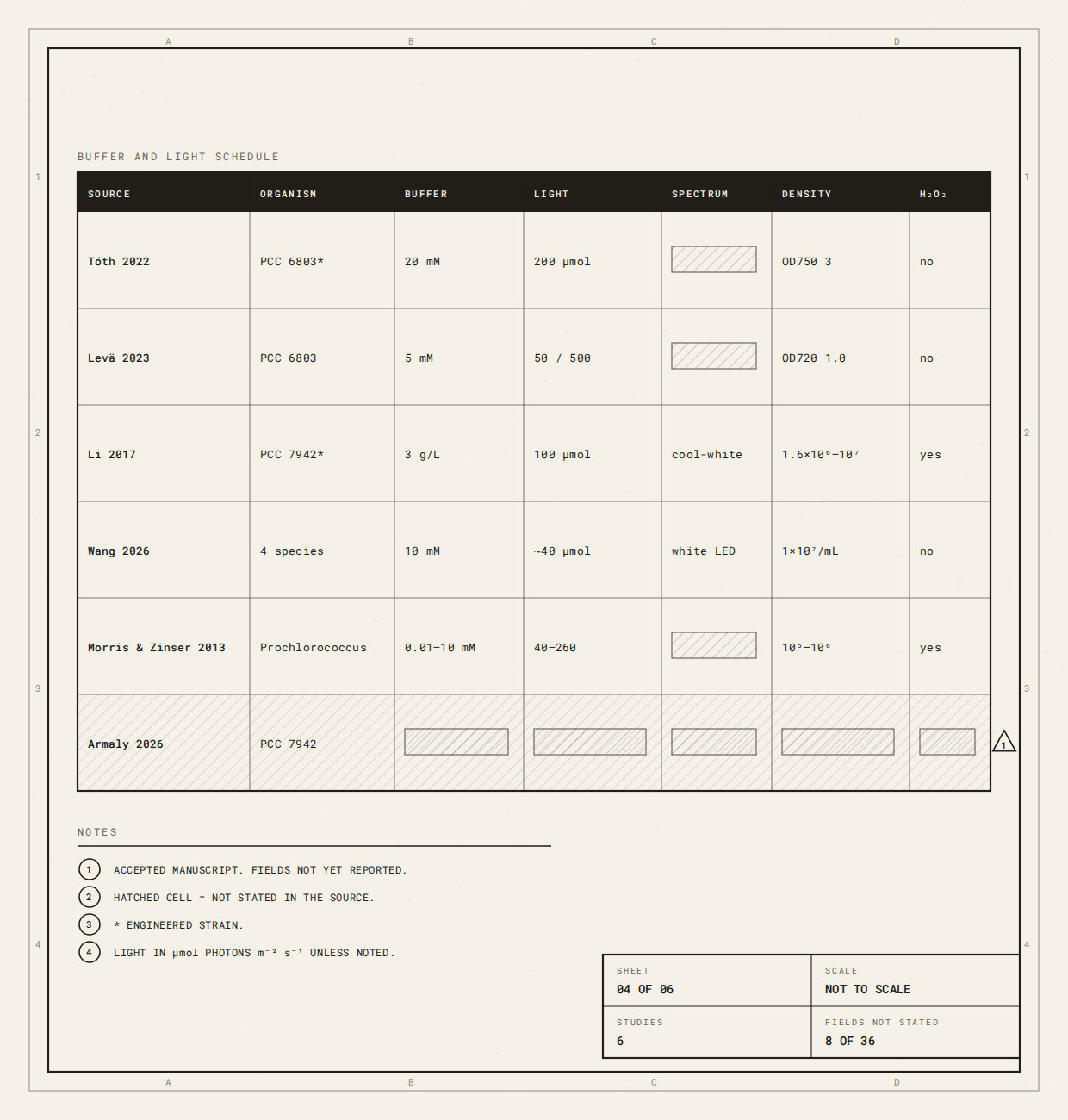
Fig. 4: What “HEPES-buffered” doesn’t tell you. Six published photosynthetic systems and the conditions that decide whether HEPES photochemistry could matter in each: organism, buffer and concentration, light intensity and spectrum, cell density, immobilisation, and whether peroxide was measured at all. The blank cells aren’t omissions from this table. They’re what the methods sections report, and one row is blank almost end to end. Compiled from the cited papers, September 2026. Read it as a map of what is stated, not a judgement of the work.
One of them describes a chain running the other way. In Levä’s scaffolds, ion exchange with the growth medium reverses part of the calcium crosslinking holding the material together. The network loosens, that changes gas exchange, and gas exchange changes carbon fixation.
The matrix that best resisted the loosening fixed the least carbon by day 5. The one that loosened kept gas exchange better, at the cost of mechanical stability. The differences weren’t statistically significant, so this is the explanation the authors propose rather than an effect they demonstrate (Levä et al., 2023).
The medium was acting on the material, and the material on the biology. This is the coupling Dawiec-Liśniewska and colleagues asked about, here with a biological consequence.
What biodesign already teaches us
Biodesign increasingly asks students and practitioners to approach living organisms as active collaborators rather than inert materials (Nerlich et al., 2026).
Nerlich, Archer and Morgado Diniz’s Living Pigments framework teaches students to prepare and modify nutrient media, manage light and gas exchange, and monitor growth through cell density estimation and growth curves. It also asks them to observe and respond to biological variability rather than treat the living system as something fully under control (Nerlich et al., 2026).
On that unit, maintenance stops being a corrective task and becomes a design condition in its own right (Nerlich et al., 2026). Keeping something alive is part of the work, not the overhead around it.
The HEPES case extends that one step further. Ask how the environment changes the organism. Then ask how it changes the things we introduced to control the organism.
That’s no departure from what the unit already does, since students modify the medium themselves. The question is what else changes when they do.
Protocols organise substances by intended role, and those labels organise attention. A hydrogel is expected to interact with cells, so its properties get investigated. A buffer is expected to reduce variation, so it fades into the background.
HEPES shows why that background deserves attention too.
When the protocol becomes machine-readable
This gets more consequential as biodesign moves towards AI-assisted experimentation and robotic fabrication.
A person reading a protocol can notice that a reagent labelled buffer might join in other chemistry. A machine-readable version is more likely to encode HEPES as pH buffer, light as photosynthetic input, cell density as biomass.
Those categories make living systems easier to manipulate, but control interference happens between them.
Imagine a robotic platform tuning a photosynthetic living material. It varies light, cell loading, thickness and geometry while HEPES stays fixed, so from the system’s perspective the buffer is controlled.
Chemically that may not be enough. Changing light changes both photosynthesis and medium photochemistry. Changing cell loading changes both growth and peroxide-removal capacity (Li et al., 2017). Changing geometry alters light penetration, and both papers above treat mass transport as something geometry decides (Dawiec-Liśniewska et al., 2026; Armaly et al., 2026).
The robot has held the amount of HEPES constant without holding its chemical consequence constant. The algorithm may still find the best-growing condition. The difficulty comes when we interpret why.
Automation doesn’t create that ambiguity. It makes it easier to reproduce at scale.
Retrieval poses a related problem. HEPES = buffer is simple to extract, and the conditional version isn’t. A database could faithfully preserve “HEPES, 20 mM, pH buffer” while losing the conditions that decide whether it’s sufficient.
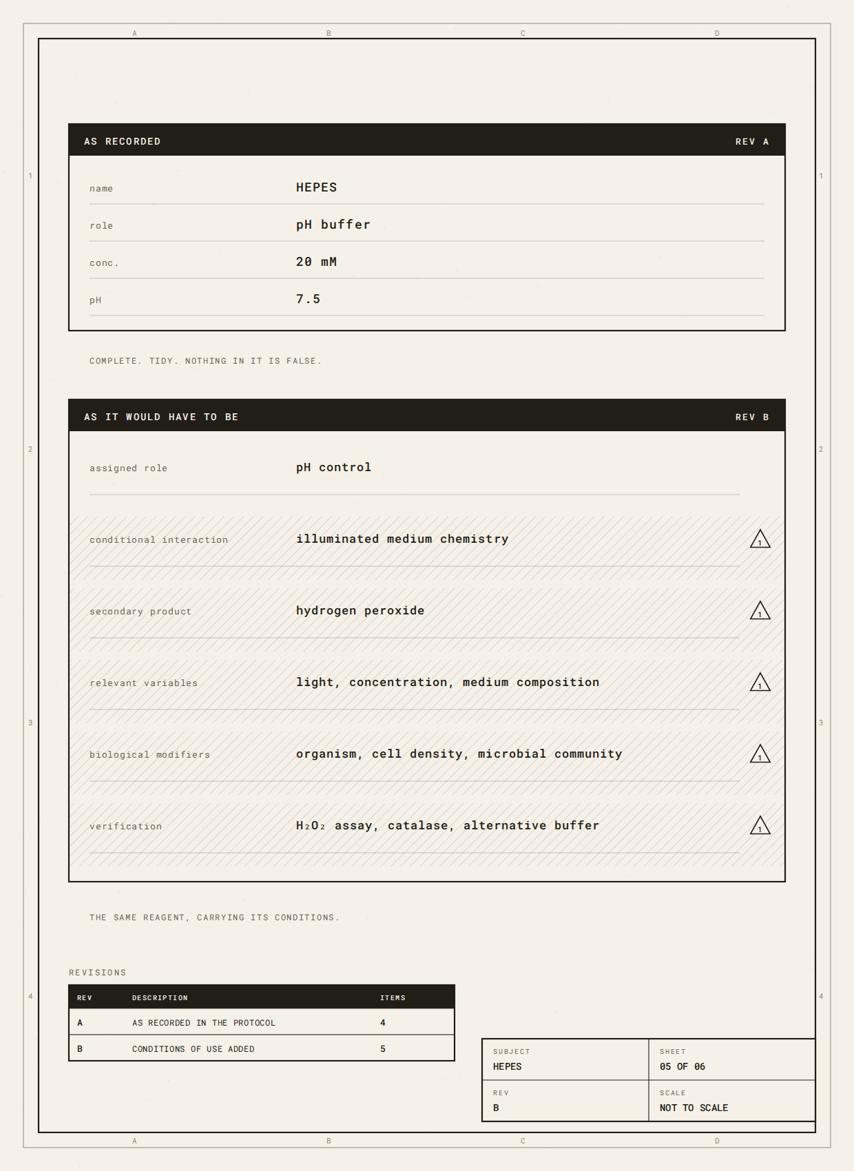
Fig. 5: Two records of the same reagent. Top, HEPES as a protocol database stores it. Nothing in it is false. Bottom, the same entry carrying the conditions that decide whether the top one is sufficient. A retrieval system can copy the top record perfectly and still move a protocol into a context where it no longer holds.
Machine-readable protocols therefore need relationships. What is this component for, and which variables change what it does?
Neutrality is not a property of the bottle
The wrong lesson would be: don’t use HEPES under light.
It works in many illuminated systems. Even in Li’s experiment, dense cultures grew strongly in the medium that failed at lower density (Li et al., 2017).
What has actually been tested, and the handful of things that shift the outcome in practice, sits on the reference page: Does HEPES produce hydrogen peroxide under light?
What changed was the configuration: light × medium chemistry × buffer concentration × cell density × organism × community.
Neutrality was an outcome of the system.
Biodesign relies on ingredients introduced to make living systems easier to work with: buffers, antibiotics, chelators, crosslinkers, inducers. Each is assigned a job. The useful question is what else becomes coupled to that control when the experiment changes around it.
When the lights come on we usually ask what happens to the organism. HEPES suggests another question. What happens to the rest of the experiment?
What to do about it
You don’t have to stop using HEPES. You have to know when the question is worth asking, and what to do if the answer is yes.
Suspect it when several of these are true together.
- —The culture is lit for long periods.
- —The buffer is an amine at millimolar concentrations.
- —The medium carries a photosensitiser such as riboflavin, added deliberately or smuggled in by a vitamin mix.
- —Cells start dilute, or the organism has thin oxidative defences.
- —The experiment runs for days rather than hours.
Any one of those is unremarkable. Together they’re the configuration in which every demonstrated case has happened.
The first check is cheap. Measure peroxide in your medium, with no cells in it, under your own light, over your own timescale. Li and colleagues ran exactly this control, and it’s the strongest evidence in their paper (Li et al., 2017; Morris & Zinser, 2013).
It costs one assay and no biology, and it tells you whether there’s anything to chase.
If there is, the levers differ in what they cost you. Changing buffer is cleanest. Tris produced no detectable peroxide in 2 independent studies, and TAPS far less than HEPES, though at 10 mM TAPS suppressed growth itself, and both differ from HEPES in buffering range, which you’ll need to check against your working pH (Liu et al., 2023; Morris & Zinser, 2013).
Catalase works quickly. It completely restored growth in one study and rescued dilute cultures at 1.25 mg/L daily in another (Morris & Zinser, 2013; Li et al., 2017). But catalase is itself photo-inactivated, so it needs topping up rather than adding once (Morris et al., 2011).
Starting denser, or co-culturing with a scavenging partner, shifts the balance from the biological side instead.
That last option is worth sitting with. In both studies where a living partner was tried against purified catalase, the partner held its own or won. Morris and colleagues found catalase clearly inferior to co-culture with the helper bacterium (Morris et al., 2011), and in Li’s dense cultures the yeast beat the enzyme outright (Li et al., 2017).
Sometimes the way to hold a system steady is to put something else living into it, rather than take a variable out.
One thing does not work. Lowering the buffer concentration is not a fix on its own. Morris and colleagues cut peroxide production by 75% and the cultures still died (Morris et al., 2011).
One is free. Keep buffered media in the dark. Media left under ordinary bench lighting for a week were lethal to Prochlorococcus cultures, and adding catalase before inoculation removed the effect completely (Morris & Zinser, 2013).
Then write it down. Record buffer identity and concentration. Light intensity and photoperiod during both cultivation and measurement. Cell density at inoculation. Medium composition. And whether peroxide was measured at all.
That last field is the one nobody fills in, and the one that tells the next person, or the next model, whether your protocol description was ever sufficient.
Members’ Teaching Notes
For personal use, to help you interrogate the Reading. Not a ready-to-teach lesson plan.
Teach the missing arrows
Convert a cultivation protocol into a causal diagram (HEPES → pH, light → photosynthesis, cell density → biomass, hydrogel → structure), then ask which arrows are missing.
Candidates: light → medium photochemistry; cell density → peroxide-removal capacity; thickness → light penetration; porosity → gas and molecular transport; community composition → extracellular chemistry.
The objective is to see that ingredient-function pairs are abstractions, not causal descriptions. Then try it on your own work: I thought changing ______ would change ______. It might also change ______ because ______.
Four questions for auditing a control
What is it supposed to control? For HEPES, pH. What else changes its behaviour? Illumination, concentration, medium chemistry. What decides whether that matters? Organism, density, community, peroxide-removal capacity. Does your design intervention change any of those?
The fourth matters most. A fabrication decision can change the conditions under which a reagent was tested.

Fig. 6: Auditing a control, 4 questions. A blank card, for printing or copying. Use it on any background component in a protocol you didn’t write.
Design the missing control
A dense culture outperforms a dilute one under the same light. What would you measure before concluding that density itself improves photosynthesis? Extracellular H₂O₂, a matched-pH alternative buffer, catalase, independent pH monitoring, a low-light comparison.
Then ask which explanations each of those removes, and which survive.
Now a published case. Levä and colleagues followed gas exchange in immobilised Synechocystis PCC 6803 for 5 days, and rates halved across every matrix type. Two explanations are offered: biological changes in the cells, or restricted mass transfer (Levä et al., 2023). The cells had been grown in BG-11 containing 5 mM HEPES, under light.
A third pathway is never raised.
That doesn’t mean it was operating. The growth light was modest, and Synechocystis is not Prochlorococcus. What the medium contributes is also less obvious than it looks: a defined salt medium produced roughly 7 times less peroxide than natural seawater, but not none, and nobody identified what in seawater drives the reaction (Morris & Zinser, 2013). Li’s BG-11-based medium produced about 25 µM under light (Li et al., 2017).
The exercise is to notice that the question was available, cheap, and not asked. What single measurement would have closed it?
Extend it into Biodesign × AI
Imagine the experiment as a machine-readable protocol: HEPES = pH buffer, light = photosynthesis, cell density = biomass. Ask what the machine doesn’t know.
A richer record might read: assigned role, pH control; conditional interaction, illuminated medium chemistry; possible secondary product, H₂O₂; relevant variables, light, concentration, medium composition; biological modifiers, organism, density, community; verification, H₂O₂ measurement, catalase, alternative buffer.
The case teaches a habit that matters more as protocols get automated. When a protocol tells you what something is for, ask what else it can do, what changes that behaviour, and whether your intervention changes those conditions.
These notes are prompts, not a classroom resource or a teaching licence. The Biodesign Academy can build a teaching pack around control interference, HEPES and photosynthetic biodesign, with structured activities, learning outcomes, diagrams, discussion material and educator notes adapted to your course, studio or lab.
Armaly, P., Shachor, G., Berger, Y., Iliassafov, L., Rosenblau, K., Kashi, Y., & Barath, S. (2026). Biodesign approaches for photosynthetic microbial applications in architecture. Biotechnology Design. doi: 10.1017/S2977905726100961.
Dawiec-Liśniewska, A., et al. (2026). Biodesign of microalgae-laden engineered living materials via 3D bioprinting: a roadmap. Biotechnology Design. doi: 10.1017/S2977905726100869.
Levä, T., et al. (2023). Mapping nanocellulose- and alginate-based photosynthetic cell factory scaffolds: interlinking porosity, wet strength, and gas exchange. Biomacromolecules, 24, 3484-3497. doi: 10.1021/acs.biomac.3c00261.
Li, T., et al. (2017). Mimicking lichens: incorporation of yeast strains together with sucrose-secreting cyanobacteria improves survival, growth, ROS removal, and lipid production in a stable mutualistic co-culture production platform. Biotechnology for Biofuels, 10, 55. doi: 10.1186/s13068-017-0736-x.
Liu, P., et al. (2023). Zwitterionic betaines over HEPES as the new generation biocompatible pH buffers for cell culture. Bioactive Materials, 24, 376-386. doi: 10.1016/j.bioactmat.2022.12.028.
Morris, J. J., Johnson, Z. I., Szul, M. J., Keller, M., & Zinser, E. R. (2011). Dependence of the cyanobacterium Prochlorococcus on hydrogen peroxide scavenging microbes for growth at the ocean’s surface. PLoS ONE, 6(2), e16805. doi: 10.1371/journal.pone.0016805.
Morris, J. J., & Zinser, E. R. (2013). Continuous hydrogen peroxide production by organic buffers in phytoplankton culture media. Journal of Phycology, 49(6), 1223-1228. doi: 10.1111/jpy.12123.
Nerlich, P., Archer, L., & Morgado Diniz, N. (2026). Teaching biodesign through algal experimentation: methods for living-system prototyping. Biotechnology Design, 4, e32, 1-15. doi: 10.1017/S2977905726100328.
Tóth, G. S., et al. (2022). Photosynthetically produced sucrose by immobilized Synechocystis sp. PCC 6803 drives biotransformation in E. coli. Biotechnology for Biofuels and Bioproducts, 15, 146. doi: 10.1186/s13068-022-02248-1.
Wang, M., et al. (2026). Co-culture of mammalian cells and photosynthetic microorganisms for oxygen supply in engineered tissues. Cell Proliferation, advance online publication, e70224. doi: 10.1111/cpr.70224.
Zigler, J. S., Jr., et al. (1985). Analysis of the cytotoxic effects of light-exposed HEPES-containing culture medium. In Vitro Cellular & Developmental Biology, 21, 282-287. doi: 10.1007/BF02620943.
From the Molecule Up, by Biodesign Academy. Read one living material with care, as a relation you are joining, then meet the next on its own terms.
