What is structural colour?
The colour produced by the physical arrangement of a material rather than by a pigment or dye.
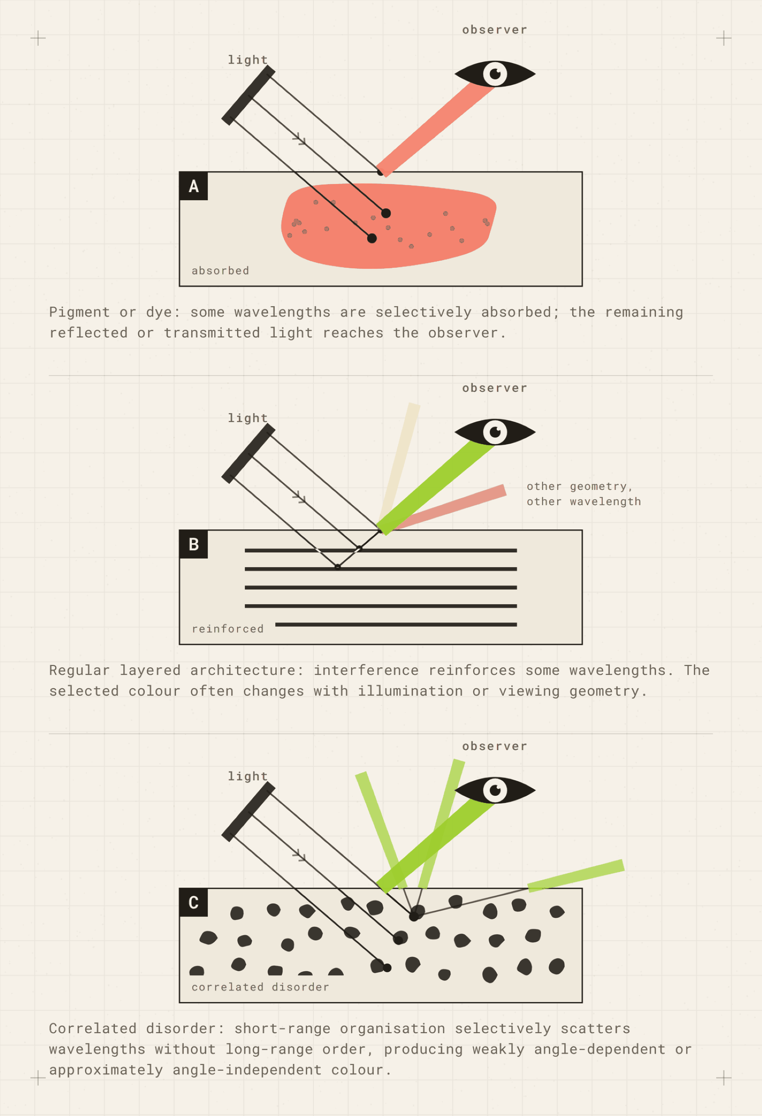
Structural colour is the colour produced by the physical arrangement of a material rather than by a pigment or dye. The arrangement reflects, redirects and scatters light so that some wavelengths reach the eye more strongly than others.
In layered structures this happens because reflected waves reinforce at some wavelengths and cancel at others.
The relevant features are often on the scale of tens to hundreds of nanometres, although larger scale structures can also shape how the colour is ultimately seen. Different structures manipulate light in different ways, so structural colour is not one single mechanism (Kinoshita et al., 2008).
This means the colour is carried by architecture. Change the spacing, orientation, shape or optical properties of that architecture and the colour can change too.
A material can also contain pigment and structural colour at the same time. The important distinction is therefore not simply whether pigment is present. It is what is doing the work of selecting the colour you see.
What is usually claimed
In design writing, structural colour is sometimes characterised as colour without pigment, although that shorthand is not universally true because structural and pigmentary colour can coexist (Yau, Klockars & Rojas, 2023).
Structural colour is also being investigated as a potentially more sustainable alternative to conventional coloration, particularly in textiles. Conventional dyeing can involve intensive water use alongside dyes, pigments and other processing requirements (Jones et al., 2020; Huang et al., 2020).
No dye does not mean no environmental cost.
Artificial structural colour can itself involve plastics, metals, nanoparticles, coatings and precisely controlled fabrication. Fabrication, flexible substrates, colour stability and durability, and industrial scale manufacturing remain engineering challenges (Huang et al., 2020).
Huang and colleagues are explicit that structural coloration may bypass conventional pigment dyeing in some applications but may never completely replace traditional dyeing (Huang et al., 2020).
And even a structural colour process does not necessarily eliminate dye. Jones and colleagues printed a structurally coloured liquid crystal layer onto polyester that had first been dyed black. The dark background absorbed transmitted light and strengthened the visible effect. The authors treated removal of that dyed substrate as future work (Jones et al., 2020).
So colour without pigment or dye can describe the mechanism producing the visible colour without describing the whole material or manufacturing process.
With a dye, colour usually travels with the colourant molecule.
With structural colour, colour travels with the architecture.
That is a different engineering challenge.
What has actually been tested
Structural colour is well established in animals, including butterfly wings, beetle shells and bird feathers (Kinoshita et al., 2008). It also occurs in plants, including the intense blue fruit of Pollia condensata (Vignolini et al., 2012).
Artificial versions have been fabricated as thin films, multilayer fibres and deliberately nanostructured surfaces, while photonic crystal structures have also been assembled on textile fibres and fabrics (Kinoshita et al., 2008; Huang et al., 2020).
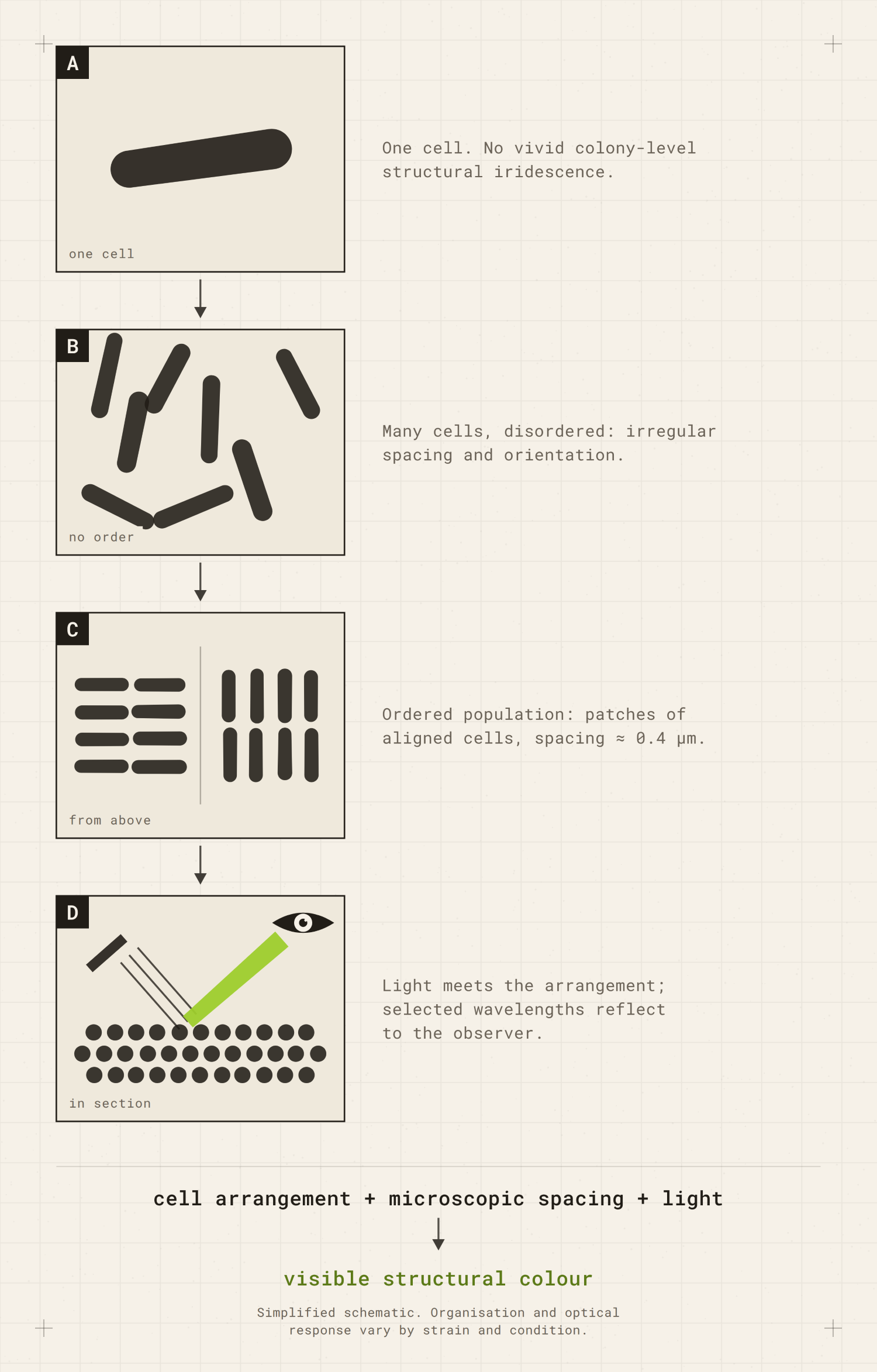
Bacterial structural colour is especially interesting because the optical architecture can be made from the arrangement of the cells themselves.
For a shorter explanation focused specifically on the bacterial case, see Why are some bacteria iridescent?
One of the best studied examples is Flavobacterium IR1.
Light as a non-destructive cell-measuring tool
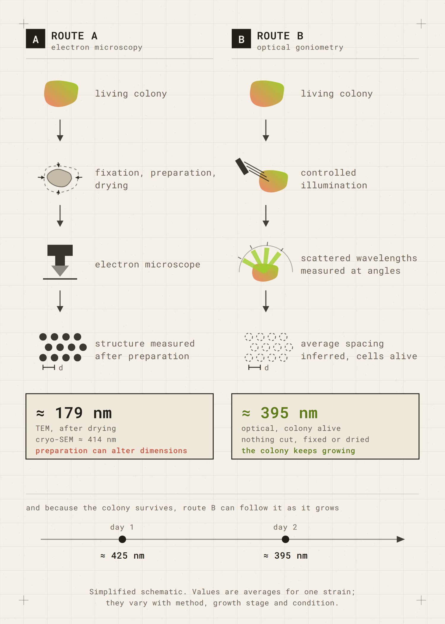
Schertel and colleagues used the optical response of living IR1 colonies to infer cell dimensions and the spacing between neighbouring cells (Schertel et al., 2020).
The important result was not simply that the colony changed colour with angle. Light became a non-destructive way of reading the organisation while the colony was still alive.
To work out how tightly the cells were packed, the researchers had to measure the gaps between them. Those gaps are a few hundred nanometres wide. A nanometre is a millionth of a millimetre, and four hundred of them is about the wavelength of violet light.
The usual tool for something that small is an electron microscope. But the standard version, transmission electron microscopy, needs the sample chemically fixed, dried out and sliced thin before it can be imaged. Prepared that way, the gaps came out at about 179 nm.
Measuring the same thing with light gave about 395 nm. More than twice as much.
The drying was the problem. The authors concluded that preparing the sample shrinks the colony, so the electron microscope was measuring accurately, but measuring something that had already shrunk. A freezing method, cryo-SEM, avoids most of that drying and gave about 414 nm, much closer to the optical figure. Their conclusion was that for averaged measurements of how these cells are packed, shining light at a living colony beat all three electron microscope methods (Schertel et al., 2020).
Because the colony stays alive, they could also measure it more than once. The same sample gave about 425 nm on day one and about 395 nm on day two. The packing had tightened while they watched.
So the colour of the colony is not something any individual cell has. It comes from where the cells sit in relation to one another.
Change the organisation and the colour changes
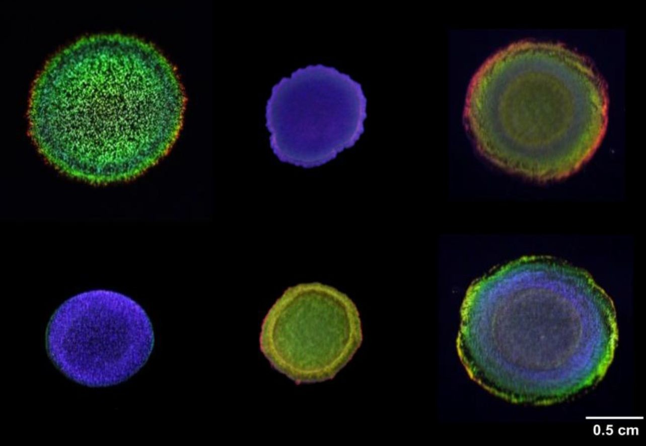
Johansen and colleagues showed that disrupting genes involved in gliding motility, cell shape, the stringent response and tRNA modification could alter colony organisation and structural colour. Their screen also identified genes associated with carbohydrate and polysaccharide related functions (Johansen et al., 2018).
One of their simplest experiments makes the point even more clearly. When they physically mixed a wild type colony with a sterile inoculation loop, the colour disappeared. It reformed within about 5 to 30 minutes as the cells reorganised (Johansen et al., 2018).
The colour could be stirred away and then rebuild itself.
Later, Escobar Doncel and colleagues used genomic information to predict a target and deliberately deleted the moeA gene. On standard medium the mutant showed a dull green blue colour, while on kappa carrageenan it produced a much stronger blue shifted appearance. The change in cell packing was inferred from the diffraction pattern rather than imaged directly (Escobar Doncel et al., 2026).
That shift from screening thousands of random mutants to targeting a predicted gene is important. The phenomenon was becoming not only observable but increasingly predictable.
The organisation is doing more than making colour
Hamidjaja and colleagues found that the ordered packing that produces IR1 structural colour was necessary for efficient predation on a surface, although not sufficient on its own (Hamidjaja et al., 2020).
Some mutants could still move but no longer formed the regular cell organisation, and they also lost the ability to efficiently degrade the prey colony. Movement alone was not enough.
Under the experimental conditions, intense angle dependent green strongly correlated with the organised state associated with efficient competition.
The colour is a readout of the organisation, not the cause of the predation.
The study does not show that structural colour evolved for predation. The authors also observed that the predation stages they examined and formation of the ordered structure were unaffected by illumination over the tested period, while leaving open the possibility that structural colour may have other functions related to light (Hamidjaja et al., 2020).
The same strain can look very different
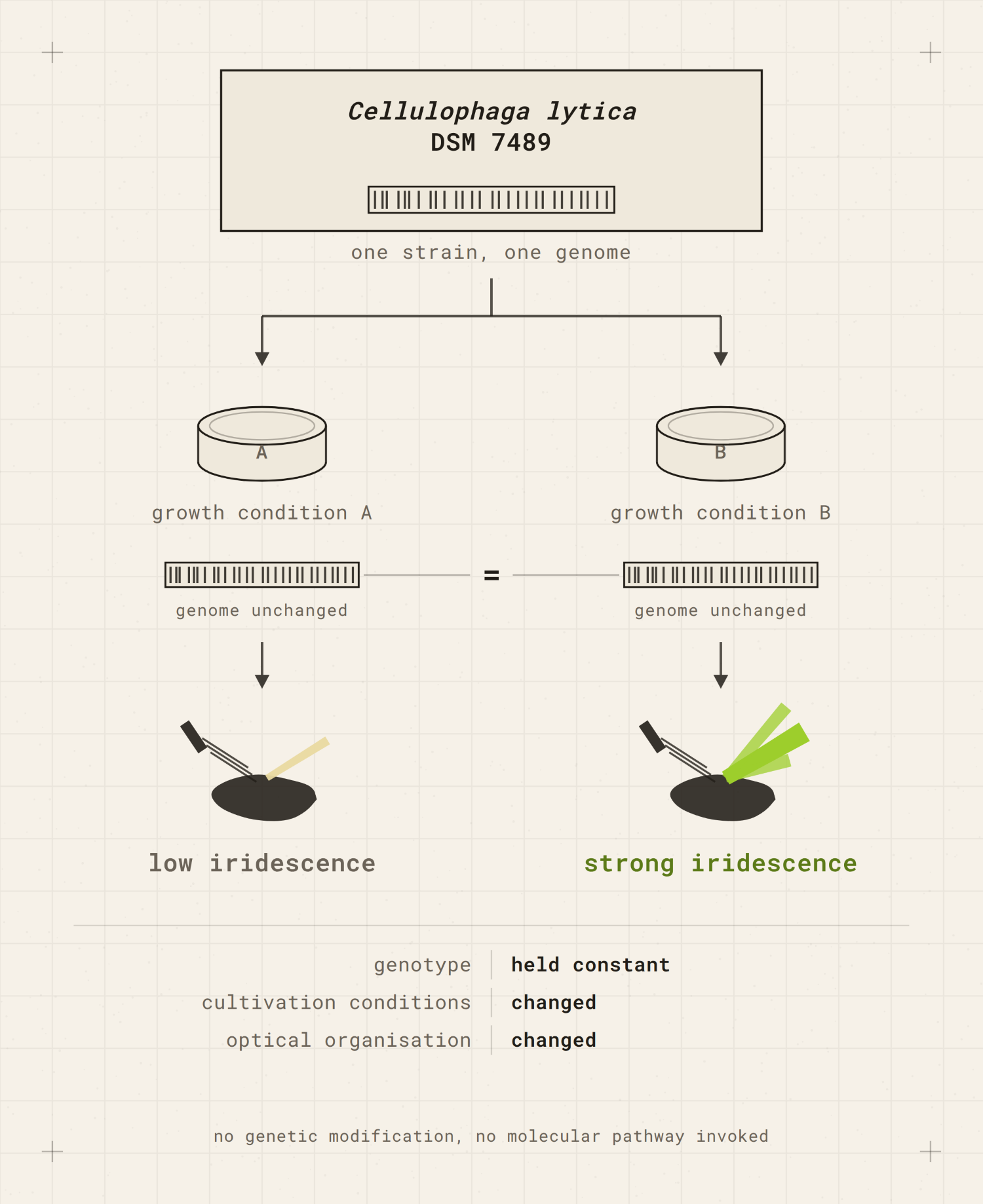
Sullivan and colleagues worked with Cellulophaga lytica DSM 7489, a strain that earlier work had used as a low iridescence comparison. Under different growth conditions, they produced strongly iridescent colonies from the same strain without genetic modification (Sullivan et al., 2023).
Same genome, different conditions, different organisation, different colour.
Sullivan and colleagues also grew biofilms on paper, polyester membranes, cotton and silk placed on nutrient agar. After fixation, pieces of biofilm could be separated from the agar in hot water and remained iridescent as floating flakes (Sullivan et al., 2023).
Paper associated biofilms lost iridescence when dried and recovered it after rehydration, and tolerated repeated drying and rehydration cycles with minimal loss of iridescence. Living biofilms could also change colour in response to changes in growth medium. Fixed biofilms did not, showing that living cells were required for that reversible response.
So the design question is not simply whether bacterial structural colour can become a material. It is what has to remain alive, organised or physically intact for the particular behaviour you want to keep.
A worked example
One of the most revealing things about IR1 is that the colour itself can be used as evidence about structure.
Schertel and colleagues shone light onto living colonies and measured the wavelengths scattered at different angles. From that optical response they could calculate how closely the cells were packed without cutting, fixing or drying the colony (Schertel et al., 2020).
You can see a simpler version of the same relationship by changing the viewing geometry.
Close to normal incidence, IR1 can appear dull and blue. Change the tilt or the direction of illumination and beyond roughly twenty degrees a much stronger green response appears. Change the geometry further and the colour and brightness shift again. Rotating the dish flat, without changing its tilt, does not produce the same effect (Schertel et al., 2020).
What changed was not the colony’s ingredients but the relationship between structure, light and observer.
That is a useful demonstration of IR1, not a universal test for structural colour. In C. lytica, for example, Sullivan and colleagues describe iridescence primarily through angle dependent changes in reflected intensity rather than necessarily a large wavelength shift (Sullivan et al., 2023).
What it is not
Not a pigment or dye mechanism
Pigments, dyes and metals produce colour mainly through interactions between light and electrons, which cause some wavelengths to be absorbed more strongly than others (Kinoshita et al., 2008).
Structural colour selects light through physical architecture. The mechanisms can also coexist in the same material.
Not the same thing as iridescence
Structural colour describes the physical origin of colour. Iridescence describes an appearance that changes with viewing or illumination geometry. That change can involve hue, brightness, or both (Kinoshita et al., 2008; Sullivan et al., 2023).
Many structural colours are iridescent. Not all are (Kinoshita et al., 2008).
The distinction matters because even iridescent structural colours do not all change in the same way. In C. lytica, Sullivan and colleagues define the bacterial iridescence mainly through an angle dependent change in peak intensity, whereas IR1 can also show pronounced changes in reflected wavelength with geometry (Sullivan et al., 2023; Schertel et al., 2020).
Not bioluminescence
Bioluminescence is light produced by a biological system. Structural colour redirects, scatters or selectively reflects light arriving from somewhere else. In darkness, the structure remains, but there is no incident light for you to see as colour.
Not automatically stable
Structural colour is sometimes described as non fading, but that is too simple.
A dry Pollia condensata fruit collected in Ghana in 1974 was still strongly blue when examined decades later. Its colour arises from a helicoidal cellulose architecture rather than a conventional blue pigment (Vignolini et al., 2012). Other structural colours can change through swelling, drying, deformation or changes in their optical surroundings.
The useful rule is that the colour lasts for as long as the material continues to provide the optical conditions needed to produce it.
Common questions
Is structural colour more sustainable than dye?
Sometimes, and not automatically.
Structural colour can reduce dependence on some conventional dyes and pigments, but producing the required microscopic architecture can introduce other materials and manufacturing demands. It may not even remove dye from the complete material system, as the black dyed polyester used by Jones and colleagues shows (Jones et al., 2020).
For textile applications, fabrication, flexible substrates, colour stability and durability, and industrial scale manufacturing remain important challenges. Huang and colleagues caution that structural coloration may never completely replace conventional textile dyeing (Huang et al., 2020).
Compare the whole process, not just the missing dye.
Can you make structural colour without living organisms?
Yes. Artificial structural colour has been made using thin films, multilayer structures, nanostructured surfaces and photonic crystals on fibres and fabrics (Kinoshita et al., 2008; Huang et al., 2020).
The distinctive proposition of structurally coloured bacteria is that the living population can participate in building the microscopic order itself.
Does structural colour fade?
It can weaken, disappear or shift if the architecture or its optical environment changes. That can happen through swelling, drying, compression, deformation, growth, mechanical damage or changes in the material surrounding the structure.
Sometimes the change is reversible. Fixed C. lytica biofilms lost iridescence when dried and recovered it after rehydration (Sullivan et al., 2023).
A better question than “will it fade?” is what has to remain physically intact for this particular structural colour to remain visible.
Biodesign gets more interesting when we stop at the mechanism long enough to ask what else is happening around it.
Subscribe to The Biodesign Academy for new definitions, in depth Library Readings, practical tools and research on the biological assumptions hidden inside materials and making.
Escobar Doncel, Á., Patinios, C., Campos, A., Walter Costa, M. B., Turkina, M. V., Murace, M., Staals, R. H. J., Vignolini, S., Dutilh, B. E. and Ingham, C. J. (2026). Deletion of the moeA gene in Flavobacterium IR1 drives structural color shift from green to blue and alters polysaccharide metabolism. eLife, 14, RP105029. DOI: 10.7554/eLife.105029.3.
Hamidjaja, R., Capoulade, J., Catón, L. and Ingham, C. J. (2020). The cell organization underlying structural colour is involved in Flavobacterium IR1 predation. The ISME Journal, 14(11), 2890 to 2900. DOI: 10.1038/s41396-020-00760-6.
Huang, M., Lu, S. G., Ren, Y., Liang, J., Lin, X. and Wang, X. (2020). Structural coloration and its application to textiles: a review. The Journal of The Textile Institute, 111(5), 756 to 764. DOI: 10.1080/00405000.2019.1663623.
Johansen, V. E., Catón, L., Hamidjaja, R., Oosterink, E., Wilts, B. D., Rasmussen, T. S., Sherlock, M. M., Ingham, C. J. and Vignolini, S. (2018). Genetic manipulation of structural color in bacterial colonies. Proceedings of the National Academy of Sciences, 115(11), 2652 to 2657. DOI: 10.1073/pnas.1716214115.
Jones, C., Wortmann, F. J., Gleeson, H. F. and Yeates, S. G. (2020). Textile materials inspired by structural colour in nature. RSC Advances, 10, 24362 to 24367. DOI: 10.1039/D0RA01326A.
Kinoshita, S., Yoshioka, S. and Miyazaki, J. (2008). Physics of structural colors. Reports on Progress in Physics, 71(7), 076401. DOI: 10.1088/0034-4885/71/7/076401.
Magkiriadou, S., Park, J.-G., Kim, Y.-S. and Manoharan, V. N. (2014). Absence of red structural color in photonic glasses, bird feathers, and certain beetles. Physical Review E, 90(6), 062302. DOI: 10.1103/PhysRevE.90.062302.
Schertel, L., van de Kerkhof, G. T., Jacucci, G., Catón, L., Ogawa, Y., Wilts, B. D., Ingham, C. J., Vignolini, S. and Johansen, V. E. (2020). Complex photonic response reveals three-dimensional self-organization of structural coloured bacterial colonies. Journal of the Royal Society Interface, 17(166), 20200196. DOI: 10.1098/rsif.2020.0196.
Sullivan, C. J., Brown, K., Hung, C. S., Tang, J. K. H., DeSimone, M., Chen, V., Lloyd, P. F., Gupta, M., Juhl, A., Crookes-Goodson, W., Vasudev, M., Dennis, P. B. and Kelley-Loughnane, N. (2023). Iridescent biofilms of Cellulophaga lytica are tunable platforms for scalable, ordered materials. Scientific Reports, 13, 13192. DOI: 10.1038/s41598-023-38797-0.
Vignolini, S., Rudall, P. J., Rowland, A. V., Reed, A., Moyroud, E., Faden, R. B., Baumberg, J. J., Glover, B. J. and Steiner, U. (2012). Pointillist structural color in Pollia fruit. Proceedings of the National Academy of Sciences, 109(39), 15712 to 15715. DOI: 10.1073/pnas.1210105109.
Yau, N. E., Klockars, K. W. and Rojas, O. J. (2023). Framing structural colour: terminology, appearance, and relevance for design. Journal of the International Colour Association, 34, 24 to 49. DOI: 10.82019/JAIC.3403.
