Why are some bacteria iridescent?
Because large numbers of cells organise into microscopic arrangements that interact with visible light.
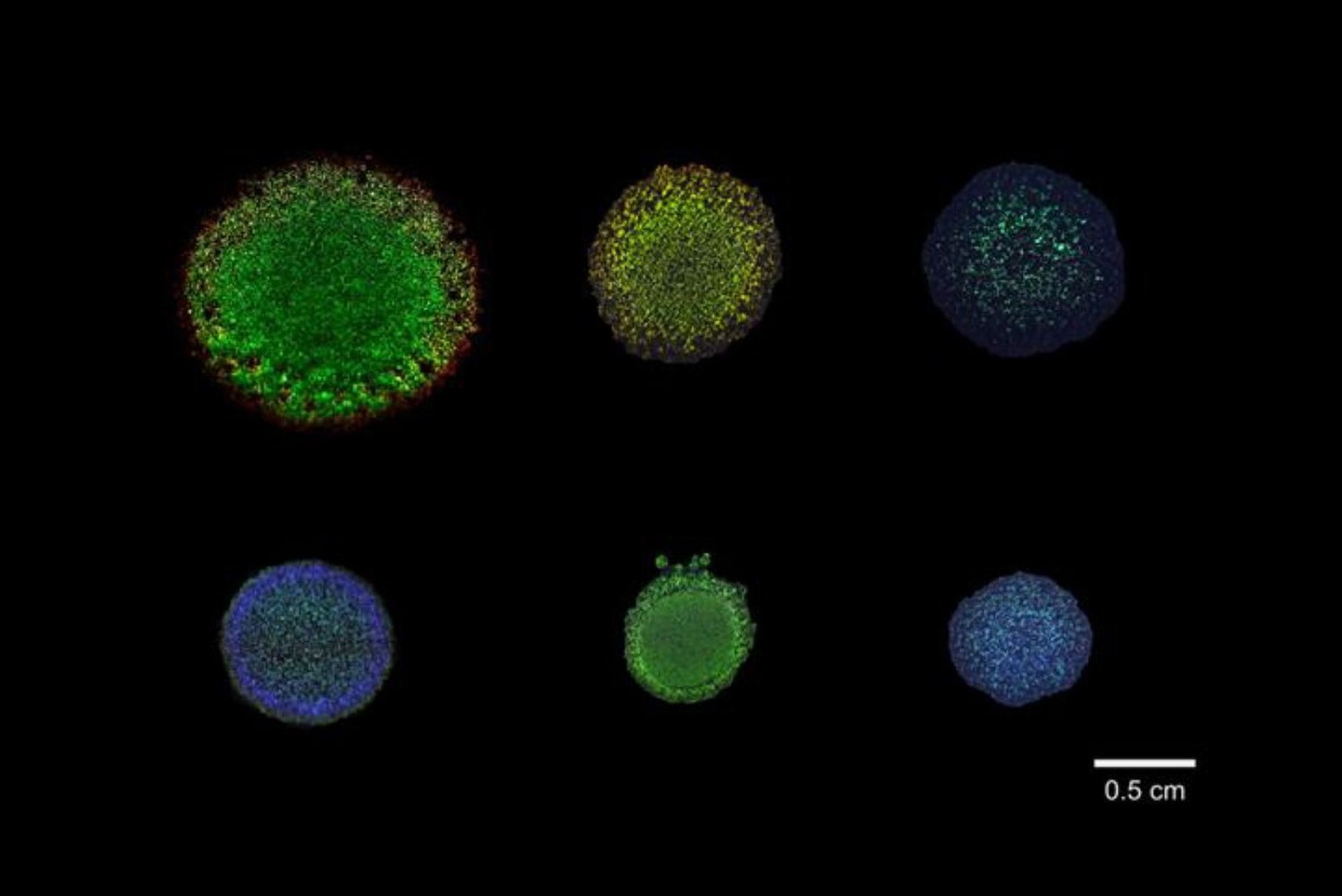
Some bacteria are iridescent because large numbers of cells organise into microscopic arrangements that interact with visible light.
The vivid colour is not simply a pigment contained inside each bacterium. It emerges when cells become organised at characteristic distances and orientations, creating an optical structure that selectively reflects or scatters particular wavelengths.
Change the spacing between cells, their orientation, the growth conditions, or the angle of illumination and observation, and the colour can change too.
This phenomenon is called structural colour, defined as the colour produced by the physical arrangement of a material rather than by a pigment or dye.
It is related to structural colours found in butterfly wings, beetle shells and bird feathers (Kinoshita et al., 2008), and in some fruits, including Pollia condensata (Vignolini et al., 2012), although the microscopic structures and optical mechanisms are not identical in every system.
Which bacteria are iridescent?
Structural colour turns up in many kinds of bacteria and in many places. In 2024, a research team sequenced 87 bacterial strains that make structural colour and 30 related strains that do not, finding the ability in at least two large branches of the bacterial family tree: the Bacteroidetes and the Proteobacteria. They then built a tool that predicts from a genome alone whether a bacterium is likely to make it, and ran it across more than 13,000 metagenomes. The ability is common in seawater and where surfaces meet air, and mostly missing from bacteria living on or inside plants and animals. Microalgae are the main exception (Zomer et al., 2024).
Two of the most studied examples are Flavobacterium IR1, a strain isolated from estuarine sediment in Rotterdam harbour, and Cellulophaga lytica, including strains that form strongly iridescent colony biofilms under particular cultivation conditions.
These bacteria typically grow as populations across a surface. Under the right conditions, very large numbers of rod shaped cells become sufficiently organised for the colony to interact with wavelengths of visible light. The result can be metallic green, blue, red or yellow appearances that change with illumination and viewing geometry.
The individual cells do not display the vivid iridescence seen across the colony. The optical effect appears at another scale.
Where does the colour come from?
Think about the difference between a pile of bricks and a brick wall. The bricks have not changed, but their arrangement has. Something similar happens with structurally coloured bacteria.
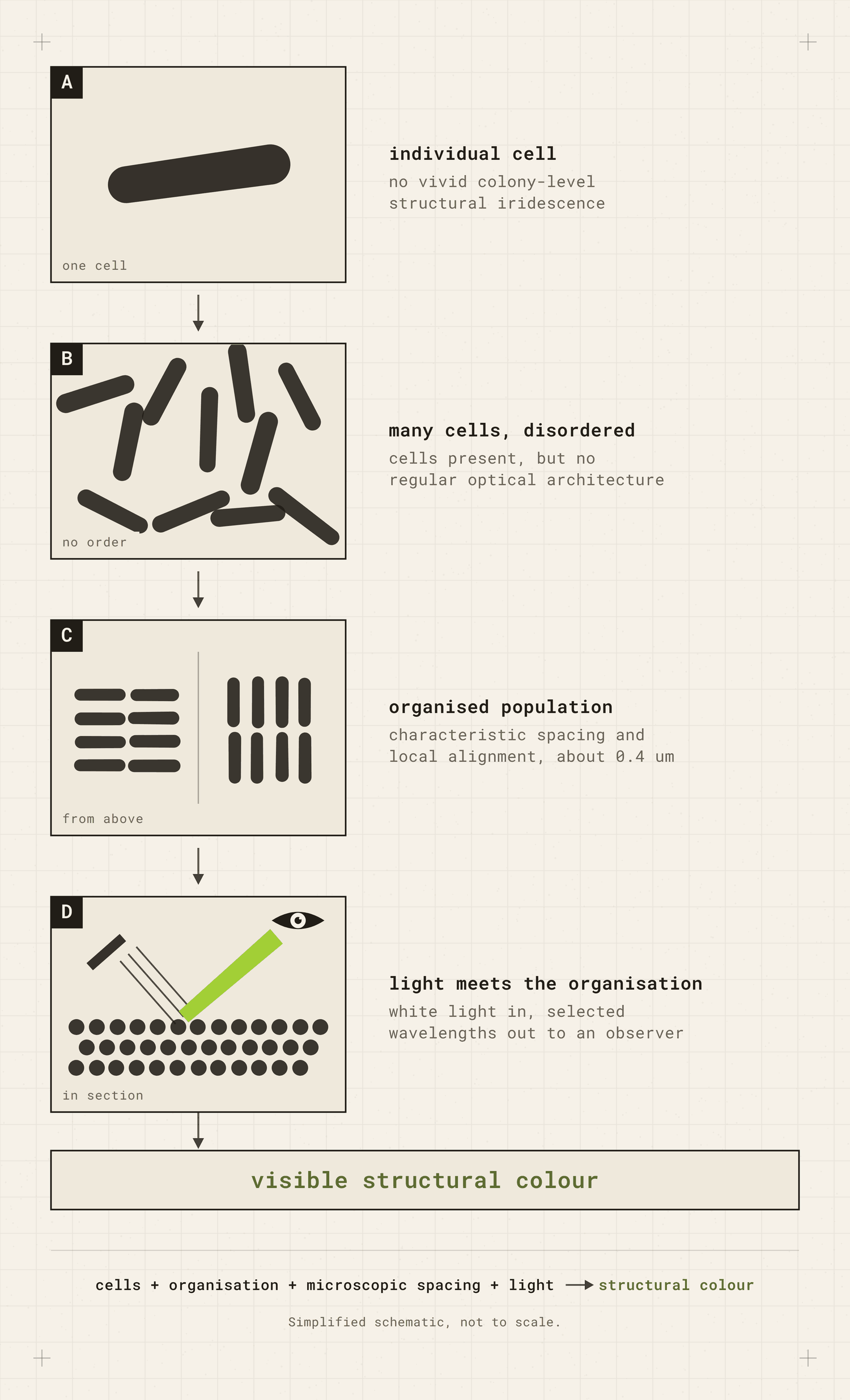
A population of randomly arranged bacterial cells does not necessarily produce a strong structural colour. When the cells become organised with sufficiently regular microscopic spacing, their collective arrangement begins to interact with light.
In Flavobacterium IR1, optical measurements have found characteristic cell spacing of roughly 400 nanometres in strongly structurally coloured colonies. That distance is comparable to the wavelengths of visible light (Schertel et al., 2020).
The cells themselves are part of the structure
Spacing and orientation are not the only physical variables that matter. The dimensions of the cells themselves can also contribute to the optical structure.
Sullivan and colleagues measured cells from differently coloured regions of the same C. lytica DSM 7489 biofilm. Cells in green regions had a mean width of about 310 nm, while cells in red regions averaged about 428 nm. They concluded that changes in morphology and cell width contributed to colour variation across the biofilm (Sullivan et al., 2023).
The cells are not simply producing different colourants. Changes in the physical components of the optical architecture contribute to which colour becomes visible. Light scattered from different parts of the organised population can then reinforce particular wavelengths more strongly than others. The observer sees the result as colour.
More accurate: a population of these bacteria can organise into a structure that produces colour.
Less accurate: this bacterium is green.
Two mechanisms in one colony
Structural colour and pigmentary colour can also occur in the same bacterial colony.
In C. lytica DSM 7489, Sullivan and colleagues observed structurally coloured bands of blue, green and red around a central region that often appeared golden-orange. They suggested that the golden-orange centre could come from the pigment zeaxanthin, which is known to occur in C. lytica. Optical measurements showed that this central region remained close to baseline reflected intensity, unlike the strongly reflecting iridescent regions around it (Sullivan et al., 2023).
A pigmented centre.
A structurally coloured surrounding biofilm.
That is useful, because the distinction is not simply between a bacterium that has pigment and one that has structural colour. The question is which mechanism is producing the particular colour you are looking at, and where.
Why does the colour change when you move?
Because structural colour depends on geometry.
With many pigments, the colour remains broadly similar when viewed from different directions because particular wavelengths are absorbed through interactions between light and electrons in the material (Kinoshita et al., 2008).
With an iridescent structure, the wavelengths reaching your eye can depend strongly on the relationship between the microscopic structure, the illumination and the observer.
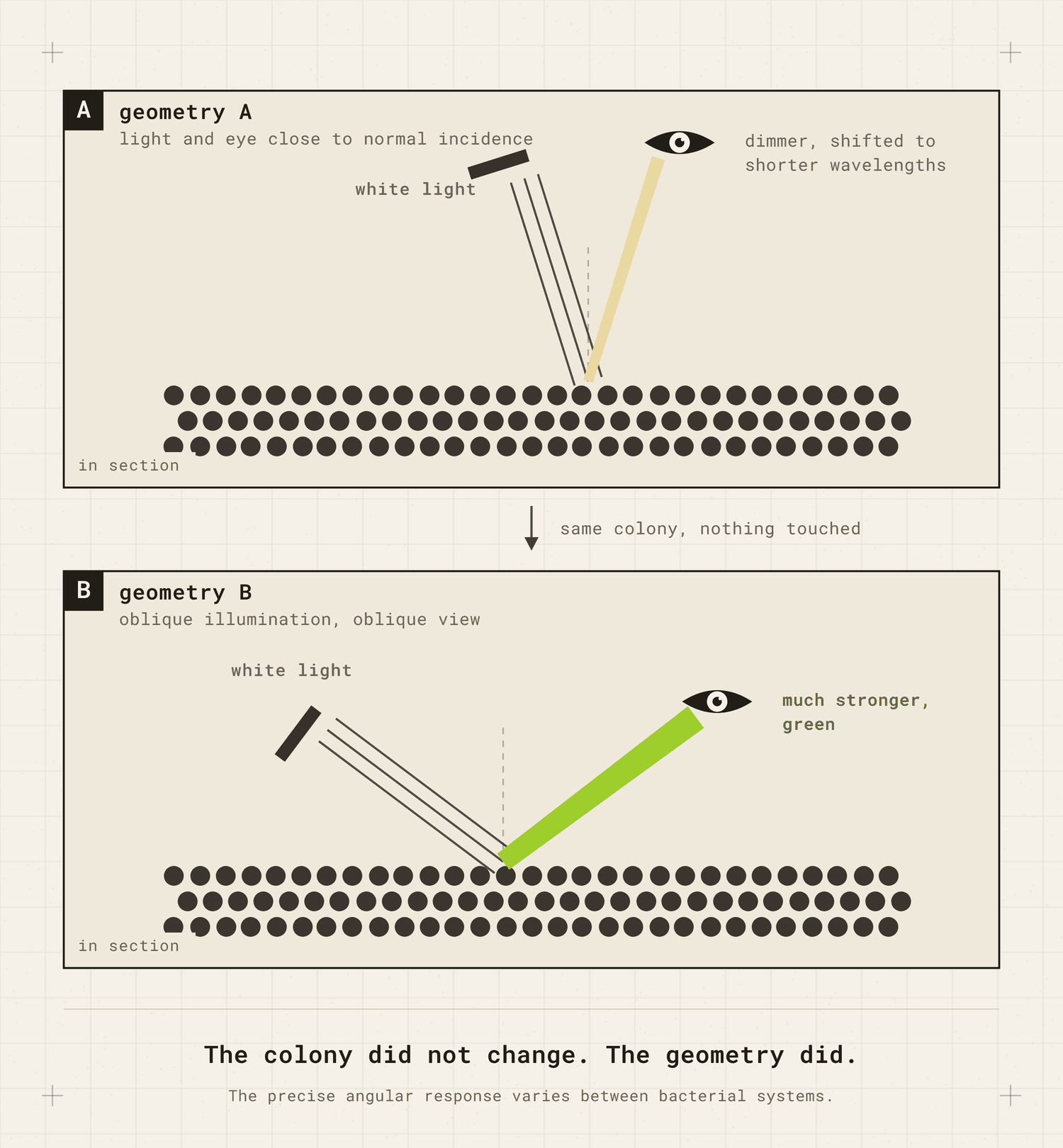
Schertel and colleagues measured this in living Flavobacterium IR1 colonies. Close to normal incidence, the colonies could appear relatively dim and blue. At other angles, a much stronger green response became visible (Schertel et al., 2020).
The colony did not change. The geometry did.
Not every iridescent bacterium behaves in exactly the same way. In Cellulophaga lytica, for example, some of the angular change is expressed particularly strongly as a change in reflected intensity rather than as a dramatic shift from one hue to another (Sullivan et al., 2023).
Iridescence therefore describes the angle dependent appearance, not one universal bacterial optical mechanism.
What makes the cells organise?
There is no single switch.
Research on Flavobacterium IR1 shows that structural colour depends on several parts of the organism’s biology, including cell movement, cell shape, population organisation and metabolism.
Johansen and colleagues screened more than 20,000 mutant colonies and found that disrupting genes involved in gliding motility, cell shape, the stringent response and tRNA modification could change or remove structural colour. They also identified genes associated with carbohydrate and polysaccharide related functions (Johansen et al., 2018).
But movement alone was not enough. Some mutant cells could still glide across the surface but failed to organise into the regular arrangements required for strong structural colour.
A colony disturbed with a wire loop
One particularly simple experiment demonstrated how directly the colour depends on organisation. The researchers physically disturbed a structurally coloured colony with a sterile inoculation loop.
The colour disappeared. Within roughly 5 to 30 minutes, it returned as the cells reorganised.
The cells had not suddenly lost and remade a green pigment. The arrangement had been disrupted and then rebuilt (Johansen et al., 2018).
Can growth conditions change bacterial iridescence?
Yes. This is one reason bacterial structural colour is particularly interesting for biodesign.
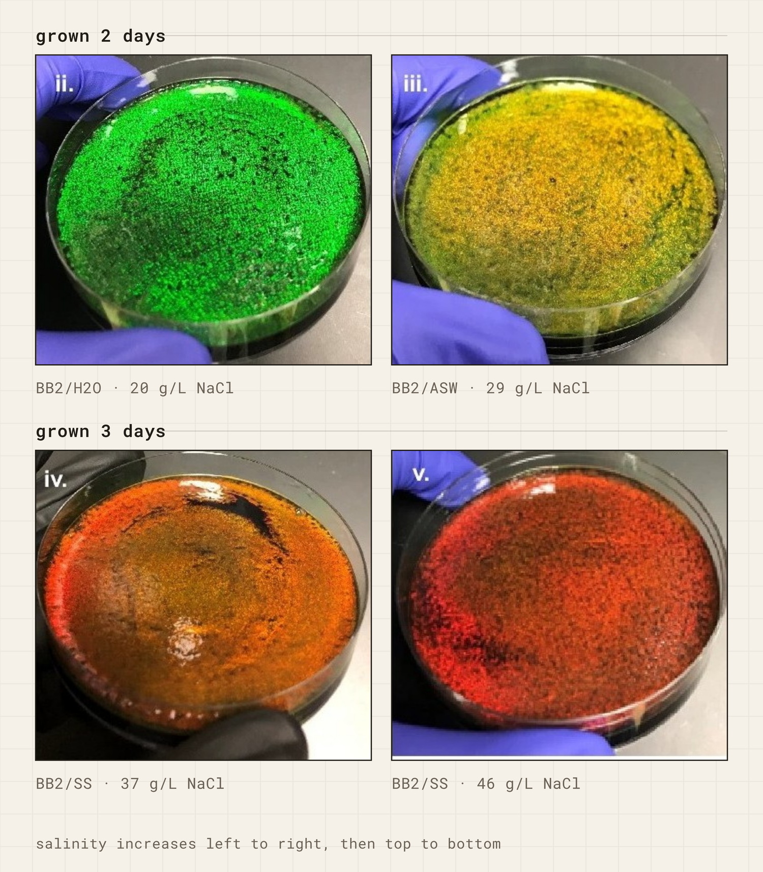
Sullivan and colleagues studied Cellulophaga lytica DSM 7489, a strain that had previously been used as a relatively low iridescence comparison. By changing cultivation conditions, they produced strongly iridescent colonies from the same strain without genetically modifying it (Sullivan et al., 2023).
The genome was the same. The conditions changed, the organisation changed, and the visible colour changed with it.
Other work with Flavobacterium IR1 has shown that polysaccharides in the growth medium can shift the colour a colony displays. On high-fucoidan agar, IR1 appears a dull red-purple rather than green (Hamidjaja et al., 2020; Escobar Doncel et al., 2026).
More recently, Escobar Doncel and colleagues deliberately deleted the moeA gene in IR1 and produced a blue shifted structural colour phenotype under particular cultivation conditions (Escobar Doncel et al., 2026).
So bacterial structural colour is influenced by both genotype and environment. It is not a fixed colour swatch attached to the name of a species or strain.
Is the colour doing anything for the bacteria?
Possibly, but this requires care. The most striking evidence comes from Flavobacterium IR1.
Hamidjaja and colleagues found that formation of the densely organised cell packing associated with structural colour was necessary for efficient predation on a surface, though not sufficient on its own (Hamidjaja et al., 2020).
The distinction matters. Some mutants could still move but could no longer maintain the organisation associated with strong structural colour, and their later predatory performance was impaired. The earliest stage of the interaction was less dependent on this organisation: a motile but disorganised mutant could still begin infiltrating and surrounding the prey colony. The organised packing became important for efficient progression of the predatory process (Hamidjaja et al., 2020).
That does not mean that green structural colour causes predation. It also does not establish that iridescence evolved because these bacteria hunt other microorganisms.
The organisation required to produce structural colour also has a biological function in facilitating predation, but that function is not itself dependent on the colony’s photonic appearance.
The predatory behaviour examined in the study, and formation of the organised structure, did not require illumination over the tested period (Hamidjaja et al., 2020). But that result should not be stretched into a claim that structural colour has no biological function related to light. The authors explicitly leave open the possibility that these highly organised colonies may have other properties connected to illumination.
The colour is a readout of the organisation, not the cause of the predation.
This is one reason these bacteria are so useful for thinking about living materials. The designer sees an optical property. The organism is doing much more.
Can bacterial structural colour be extracted like a dye?
Not in the same sense. A pigment is a substance that can sometimes be separated from the organism that produced it and applied somewhere else. With bacterial structural colour, what produces the colour is physical organisation.
Destroy that organisation and the colour can disappear. That does not mean the optical structure must always remain attached to the agar on which it was grown.
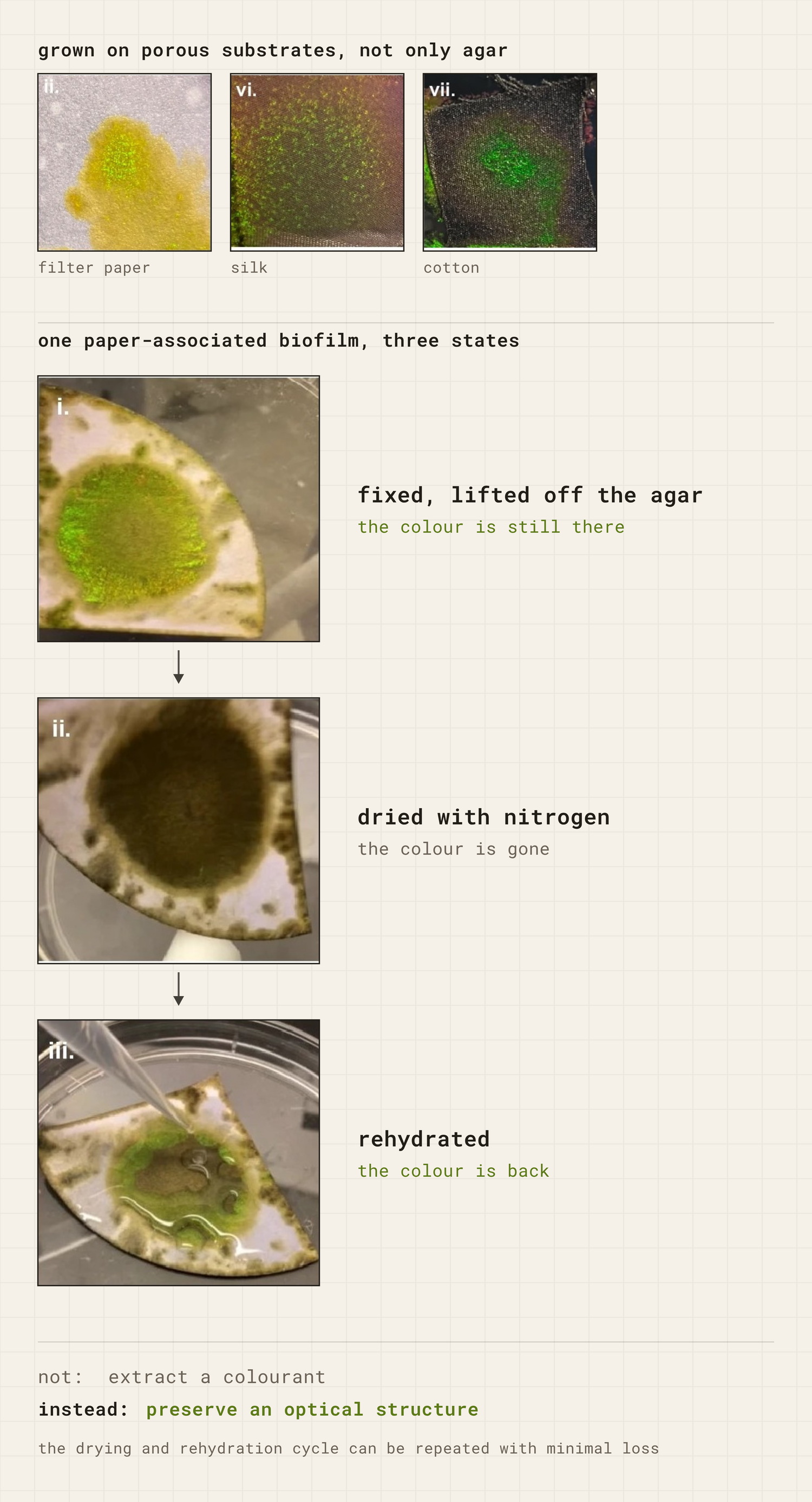
Sullivan and colleagues chemically fixed structurally coloured C. lytica biofilms with glutaraldehyde, then separated them from the agar in a hot water bath. The pieces floated free as flakes and stayed iridescent in water. Dried, they lost the iridescence and got it back on rehydration, through repeated cycles (Sullivan et al., 2023).
They also grew iridescent biofilms on materials including paper, cotton, silk and polyester membranes placed on nutrient agar (Sullivan et al., 2023).
Not: extract a colourant.
Instead: preserve the optical structure and the conditions it needs.
What is being preserved is still an optical structure, not an extracted colour molecule. That creates a different design challenge: how do you preserve the arrangement and conditions that produce the colour?
Common questions
Are iridescent bacteria bioluminescent?
No. Bioluminescent organisms produce light through biochemical reactions. Iridescent bacteria do not need to produce visible light themselves. Their microscopic organisation modifies light arriving from an external source.
Put the colony in darkness and there is no visible iridescence, because there is no incident light for the structure to redirect towards your eye. The organisation can still be present.
Are individual bacterial cells iridescent?
Not in the vivid colony level sense described here. The striking structural colour appears when many cells become organised into an optical structure.
This distinction matters because saying an iridescent bacterium is convenient shorthand, but it can hide the scale at which the phenomenon actually occurs. The colour belongs to a relationship among many cells, their environment and light.
Can you deliberately grow iridescent bacteria?
Yes, under suitable laboratory conditions. Researchers have repeatedly cultured structurally coloured Flavobacterium and Cellulophaga colonies and changed their optical appearance through variables including growth medium, surface conditions, colony age and genetic modification.
But growing a bacterium and reliably engineering a particular colour are not the same problem. The output depends on the organisation of a living population, and that organisation can change with time and environmental conditions.
Cultivation is not the preparation stage before colour appears. The cultivation conditions are part of the colour system.
Why bacterial iridescence matters for biodesign
Bacterial structural colour challenges a very familiar way of describing biological materials.
Usually: organism produces property. A bacterium produces pigment. A fungus produces mycelium. A bacterium produces structural colour.
More accurately: organism, movement, growth, environment, collective organisation and light together produce the visible property.
The difference matters. If you treat structural colour as something the bacterium simply possesses, changes in medium, hydration, colony age or cell organisation look like unwanted variability.
If you understand the colour as an emergent property of those relationships, those same variables become part of the material itself.
That is why these colonies are useful far beyond the question of whether bacteria might someday replace textile dyes. They show what can disappear when a living process is compressed into a material property.
Escobar Doncel, Á., Patinios, C., Campos, A., Walter Costa, M. B., Turkina, M. V., Murace, M., Staals, R. H. J., Vignolini, S., Dutilh, B. E. and Ingham, C. J. (2026). Deletion of the moeA gene in Flavobacterium IR1 drives structural color shift from green to blue and alters polysaccharide metabolism. eLife, 14, RP105029. DOI: 10.7554/eLife.105029.3.
Hamidjaja, R., Capoulade, J., Catón, L. and Ingham, C. J. (2020). The cell organization underlying structural colour is involved in Flavobacterium IR1 predation. The ISME Journal, 14(11), 2890 to 2900. DOI: 10.1038/s41396-020-00760-6.
Johansen, V. E., Catón, L., Hamidjaja, R., Oosterink, E., Wilts, B. D., Rasmussen, T. S., Sherlock, M. M., Ingham, C. J. and Vignolini, S. (2018). Genetic manipulation of structural color in bacterial colonies. Proceedings of the National Academy of Sciences, 115(11), 2652 to 2657. DOI: 10.1073/pnas.1716214115.
Kinoshita, S., Yoshioka, S. and Miyazaki, J. (2008). Physics of structural colors. Reports on Progress in Physics, 71(7), 076401. DOI: 10.1088/0034-4885/71/7/076401.
Schertel, L., van de Kerkhof, G. T., Jacucci, G., Catón, L., Ogawa, Y., Wilts, B. D., Ingham, C. J., Vignolini, S. and Johansen, V. E. (2020). Complex photonic response reveals three-dimensional self-organization of structural coloured bacterial colonies. Journal of the Royal Society Interface, 17(166), 20200196. DOI: 10.1098/rsif.2020.0196.
Sullivan, C. J., Brown, K., Hung, C. S., Tang, J. K. H., DeSimone, M., Chen, V., Lloyd, P. F., Gupta, M., Juhl, A., Crookes-Goodson, W., Vasudev, M., Dennis, P. B. and Kelley-Loughnane, N. (2023). Iridescent biofilms of Cellulophaga lytica are tunable platforms for scalable, ordered materials. Scientific Reports, 13, 13192. DOI: 10.1038/s41598-023-38797-0.
Vignolini, S., Rudall, P. J., Rowland, A. V., Reed, A., Moyroud, E., Faden, R. B., Baumberg, J. J., Glover, B. J. and Steiner, U. (2012). Pointillist structural color in Pollia fruit. Proceedings of the National Academy of Sciences, 109(39), 15712 to 15715. DOI: 10.1073/pnas.1210105109.
Zomer, A., Ingham, C. J., von Meijenfeldt, F. A. B., Escobar Doncel, Á., van de Kerkhof, G. T., Hamidjaja, R., Schouten, S., Schertel, L., Müller, K. H., Catón, L., Hahnke, R. L., Bolhuis, H., Vignolini, S. and Dutilh, B. E. (2024). Structural color in the bacterial domain: The ecogenomics of a 2-dimensional optical phenotype. Proceedings of the National Academy of Sciences, 121(29), e2309757121. DOI: 10.1073/pnas.2309757121.
