The protein that makes its own colour
GFP glows green without a dye. Three letters of its own chain become the colour, and one change turns it blue.
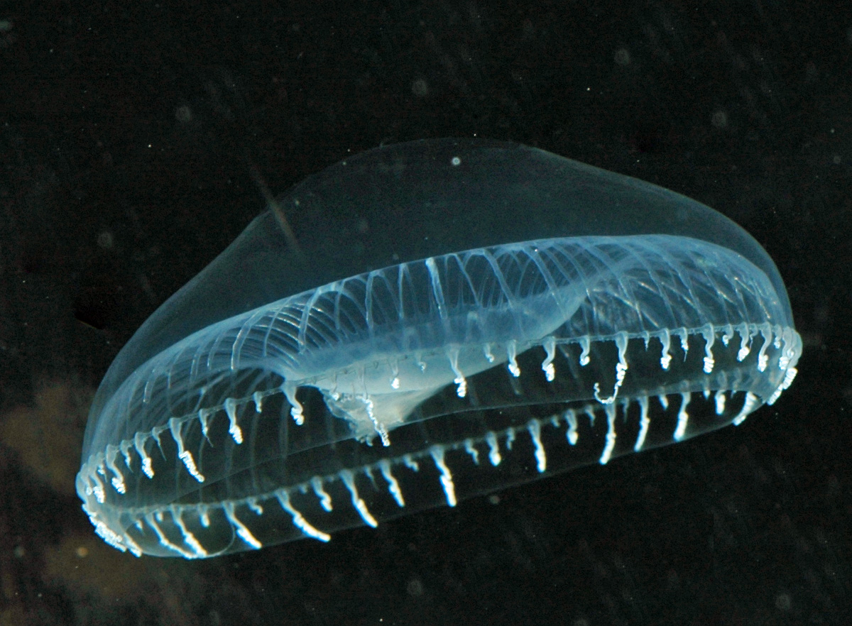
Fig. 1. Aequorea victoria, the jellyfish where GFP was found. In ordinary light it looks clear. Its green glow shows in the dark, when it flashes. Photo: Sierra Blakely, via Wikimedia Commons.
A small jellyfish in the North Pacific, Aequorea victoria, glows green at its edges. The green comes from a protein: green fluorescent protein, or GFP.
GFP is a chain of 238 amino acids, each with a one-letter name. The chain folds into a barrel of eleven strands. Deep inside sit three letters, S, Y and G, at positions 65, 66 and 67.
The protein makes its own colour
Most coloured proteins pick up a coloured molecule made somewhere else. GFP doesn’t. Once the barrel has folded, those three letters bend, join into a ring and react with oxygen. They become the chromophore, the part that catches light.
Shine blue or ultraviolet light on GFP and it gives back green light. That’s fluorescence: one colour of light in, another colour out. The barrel holds the chromophore still and shields it from water. Unfolded, GFP doesn’t glow.
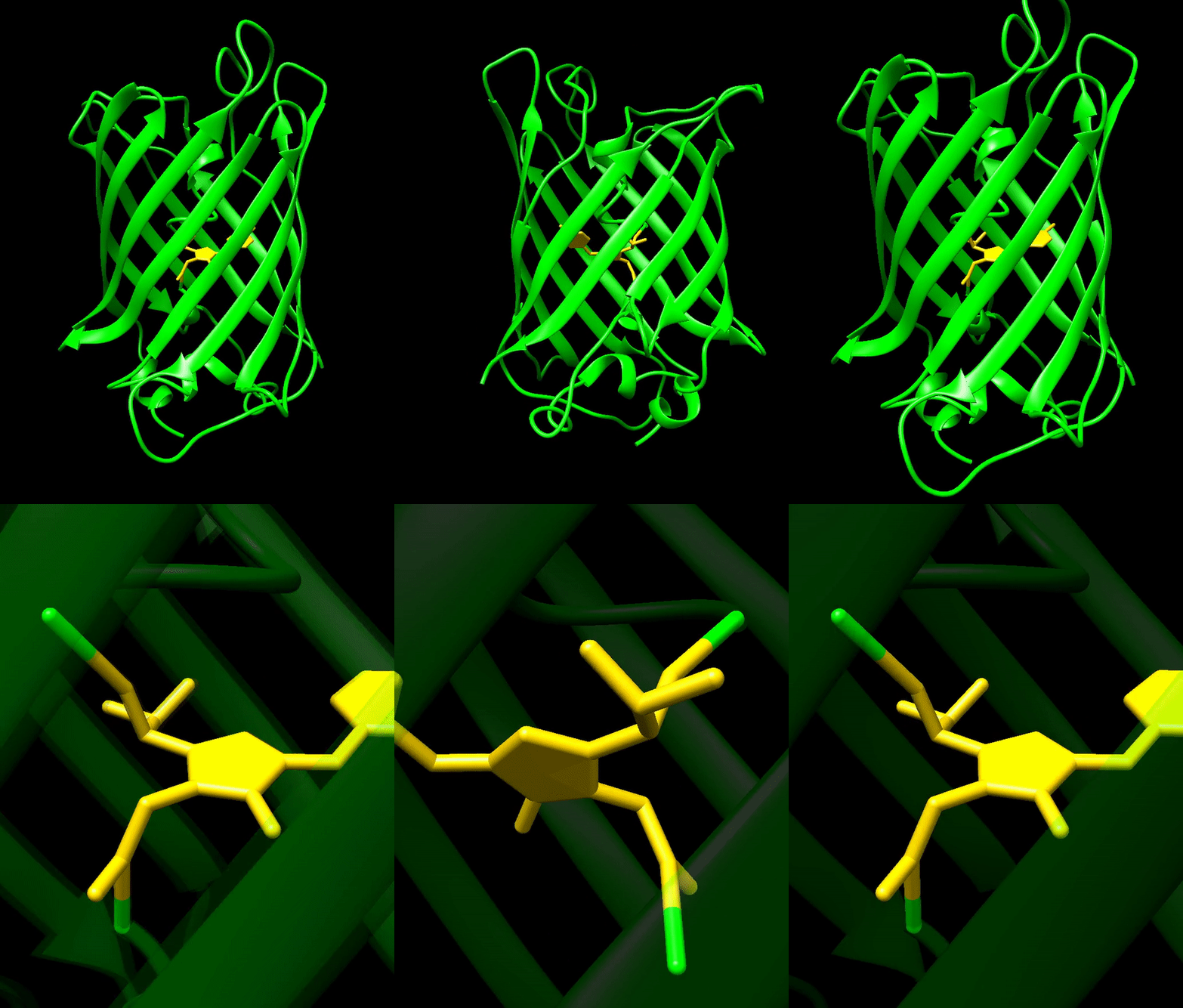
Fig. 2. Green fluorescent protein (GFP). The protein folds into a barrel of strands. The chromophore, shown in yellow, sits inside the barrel. It forms from three of the protein’s own amino acids, and it is the part that glows. Animation: Erik A. Rodriguez (Wikimedia Commons user Erin Rod), made with UCSF Chimera from PDB 1EMA. Via Wikimedia Commons, licensed under CC BY-SA 4.0.
One letter changes the colour
Change letter 66 from Y (tyrosine) to H (histidine) and the glow turns blue. Change it to W (tryptophan) and it turns cyan. Each change alters the ring inside the barrel. From the outside, the protein looks almost the same.
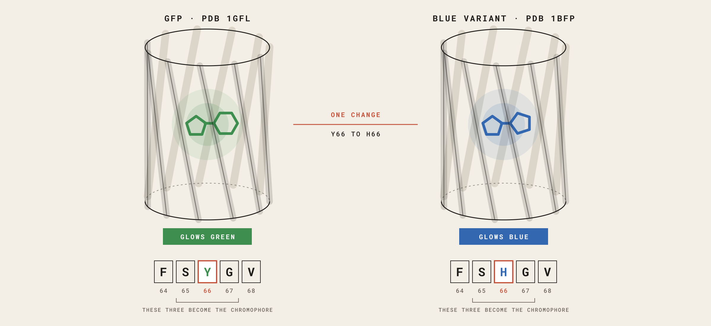
Fig. 3. Green GFP (left, PDB 1GFL) and its blue variant (right, PDB 1BFP). At position 66, Y (tyrosine) becomes H (histidine). This swaps the ring inside the barrel, and the glow turns from green to blue. The barrel barely changes. Schematic illustration by Biodesign Academy.
Glowing is not the same as coloured
Corals carry relatives of GFP. Some glow. Others, called chromoproteins, soak up light and show a strong colour in plain daylight.
The line between the two is not sharp. Every coloured protein soaks up some light. A fluorescent protein sends part of it back out as a new colour. A chromoprotein turns it into a little heat. Some fluorescent proteins soak up so much daylight that you see their colour without a lamp. Red fluorescent protein is one: the bacteria in Fig. 4 are pink in ordinary room light.
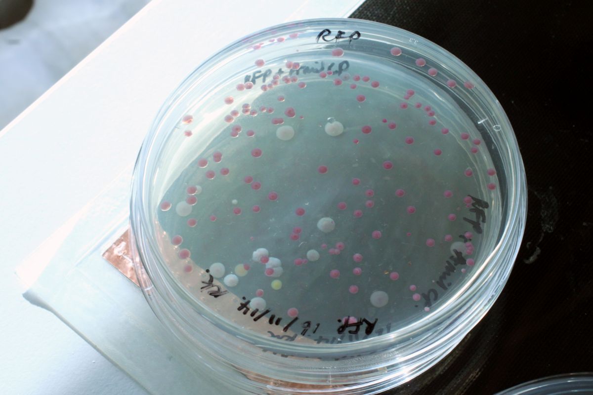
Fig. 4. Bacteria grown on a plate, photographed in ordinary room light. Most colonies are pink because their cells make red fluorescent protein (RFP), a coral relative of GFP. The few pale colonies are most likely cells that are not making the protein. Photo: Raphael Kim, 2014.
What a model can’t show
Open GFP in the AlphaFold Database and you get a good barrel. You don’t get the colour. The model shows S, Y and G as three ordinary letters, not the ring they become after folding and oxygen.
The colour lives in chemistry that happens after the fold. A structure tells you shape, not what colour comes out. That’s the judgement no model makes for you.
For designers
In GFP, colour is a recipe written into the chain. Changing one letter is a design decision with a colour as its result. But only making the protein tells you which colour you’ll get.
The full Reading, in the Library
For library members: what the AlphaFold scores do and don’t tell you about brightness, design moves for working with protein colour, and teaching notes with a “lamp on, lamp off” activity for the classroom.
AI-Native Biodesign: With Proteins is a six-week online course, 5 November to 10 December. No biology, coding or lab needed. Each week you take one step on your own project. Founding prices hold until 25 October.
RCSB Protein Data Bank. Entry 1GFL, green fluorescent protein from Aequorea victoria. rcsb.org/structure/1GFL
RCSB Protein Data Bank. Entry 1BFP, blue variant of GFP (Wachter et al. 1997). rcsb.org/structure/1BFP
AlphaFold Protein Structure Database. Predicted model for UniProt P42212. alphafold.ebi.ac.uk/entry/P42212
